No part of this publication may be reproduced, stored in a retrieval system, distributed, transmitted, translated, republished, or made available in any form or by any means without prior written permission from the copyright holder, except for brief quotations used for scholarly criticism, review, teaching, or other uses permitted by applicable law.
Medical & Educational Disclaimer
This e-book is intended for medical education and professional reference. It does not constitute personal medical advice and is not a substitute for independent clinical judgment, diagnosis, treatment planning, or consultation of current professional guidelines. Patient care must always be individualized according to the clinical circumstances, current evidence, applicable standards of care, and relevant drug, implant, or device information.
Medical knowledge evolves continuously. Although reasonable care has been taken in preparing this material, readers should verify important clinical information against current authoritative sources before applying it in practice.
Third-Party Materials, Figures & Media
Figures, photographs, illustrations, animations, video material, trademarks, and other third-party content remain subject to the rights, licenses, permissions, and attribution requirements of their respective owners. Where third-party or licensed material is used, the applicable source and credit should be identified with the individual item or in its accompanying figure credit. Nothing in this copyright notice overrides a separate third-party license or permission.
Permissions & Academic Use
Permission is required for republication, commercial reuse, translation, adaptation, or substantial reproduction of original material from this e-book unless the proposed use is otherwise permitted by law. When quoting or referring to the work in scholarly writing, readers should provide an appropriate citation to the e-book and, where relevant, to the primary scientific sources cited within it.
Rights notice: Except where otherwise credited, this notice applies to the original written content and compilation of this e-book. Individual third-party materials retain their own copyright and licensing status.
Table of Contents
1
Anatomy and Biomechanics of the Rotator Cuff
Osteology (Scapula, Clavicle, Humerus)
Functional Myology & Rotator Cable Concept
Vascular Critical Zone & Neural Pathways
Section 5 — Periarticular Bursae of the Shoulder
Section 6 — Shoulder Joint Articulations
Section 7 — Rotator Cuff Biomechanics
2
Shoulder Impingement Syndromes
Section 1 — Subacromial Impingement Syndrome
Section 2 — Internal Impingement
Section 3 — Coracoid Impingement
3
Rotator Cuff Tears
Section 1 — Partial Rotator Cuff Tear
Section 2 — Total Rotator Cuff Tear
4
Biceps Pathology
Section 1 — Biceps Tear
Section 2 — Biceps Subluxation
Clinical Teaching Case — Bodybuilder with Biceps Subluxation
5
Acromioclavicular Joint Diseases
Section 1 — Dislocations
Section 2 — AC Joint Inflammation
Clinical Teaching Case — Idiopathic AC Joint Inflammation
6
Shoulder Calcification
Uhthoff Pathophysiological Phases
US-Guided Needle Barbotage & Surgical Evacuation
7
Frozen Shoulder (Adhesive Capsulitis)
Inflammatory Synovitis & Capsular Contracture
Hydrodilatation & 360° Capsular Release
8
Shoulder Instability
Section 1 — Anterior Shoulder Instability
Bankart, Hill-Sachs & Glenoid Bone Loss
Remplissage vs Latarjet — Interactive Decision Guide
Clinical Case — Anterior Wall Failure & Posterior Subluxation
Interactive Case — Posterior Shoulder Fracture-Dislocation
1
Anatomy and Biomechanics of the Rotator Cuff Matsen Ch. 1 / Burkhart et al.
From a clinical standpoint, the shoulder gains its remarkable range of motion by accepting relatively little bony constraint. The glenoid is shallow in relation to the much larger humeral head, so stability cannot depend on bone shape alone. Instead, normal joint control relies on the combined contribution of the capsulolabral and ligamentous restraints, coordinated muscle forces, and preserved osseous anatomy [1]. Dynamic compression from the rotator cuff helps keep the humeral head centered on the glenoid during motion, complementing the static restraints and allowing mobility without sacrificing functional stability [26][28].
Clinically, shoulder motion is described in six principal directions. Flexion elevates the arm anteriorly, whereas extension carries it posteriorly. Abduction moves the arm away from the trunk, while adduction returns it toward the body. Internal rotation rotates the humerus medially and external rotation rotates it laterally. These movements are best understood as motions of the shoulder complex rather than of the glenohumeral articulation in isolation, because coordinated scapular and clavicular motion contributes progressively as the arm is elevated [1][5].
Range of motion should therefore be interpreted as a clinical spectrum rather than as a single fixed value. In a large community-based cohort of adults without shoulder pain or stiffness, mean active right-shoulder flexion was approximately 159–162° and mean active abduction approximately 150–152°, with both measures declining with age; external rotation also showed age-related variation [45]. Extension and adduction are assessed as smaller posterior and medial arcs, while internal- and external-rotation measurements are particularly dependent on the starting position and examination technique. All six directions—flexion, extension, abduction, adduction, internal rotation, and external rotation—are routinely quantified during clinical shoulder range-of-motion assessment [46]. These movements can be viewed directly in Figure 1.1 — General Movements of the Shoulder.
Figure 1.1 — General Movements of the Shoulder
Interactive three-dimensional demonstration of the principal shoulder movements: flexion, extension, abduction, adduction, internal rotation, and external rotation. Select a movement, choose the right or left shoulder, scrub through the range, and drag the model to inspect the anatomy from different viewpoints.
Interactive educational 3D anatomical module integrated into this eBook. A darker warm-neutral stage, enhanced studio/rim lighting, and optional teaching overlays preserve anatomical clarity while keeping the resting view uncluttered.
Osteological Foundations
A practical understanding of shoulder disease begins with three bones that shape the shoulder complex: the scapula, clavicle, and proximal humerus.
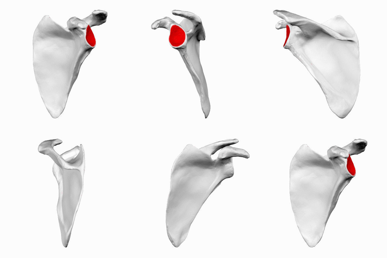
The Scapula provides the mobile base on which much of shoulder function depends [1]. Important landmarks include the glenoid cavity, which has been described as having slight retroversion and superior tilt [17]; the coracoid process, which gives attachment to several muscles and the coracoclavicular ligaments [18]; and the spine of the scapula, which continues laterally into the acromion[1]. The shape of the acromion also influences the available space beneath it [2].
A
B
C
D
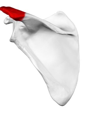
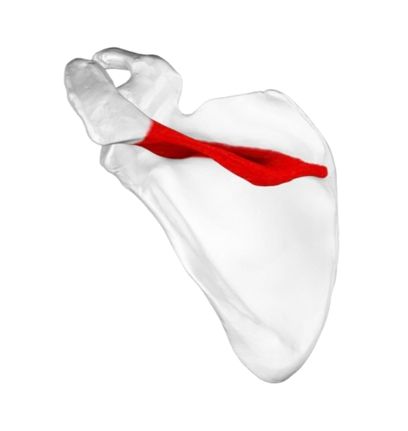
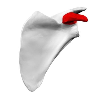
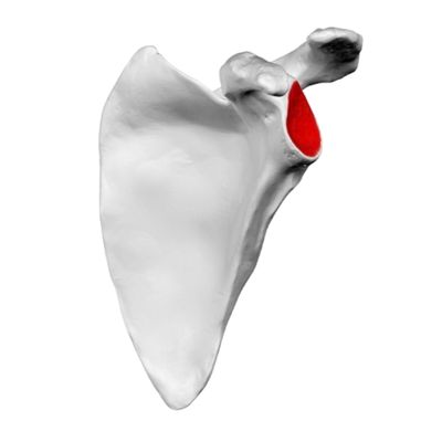
Figure 1.2 — Anatomic Features of the Human Scapula
Anatomical views illustrating key scapular landmarks highlighted in red:
(A) Acromion process of the scapula (posterior view) [8].
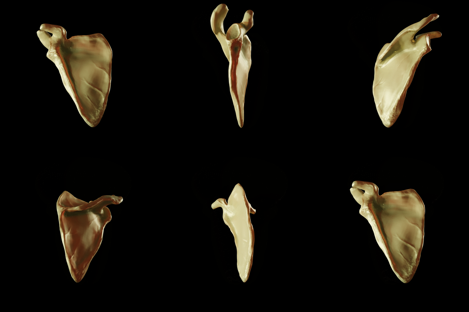
Figure 1.2A — 3D Rotation of the Human Scapula
Computer-generated 3D rotational view of the human scapula demonstrating its major anatomical landmarks, including the acromion, coracoid process, glenoid fossa, supraspinous and infraspinous fossae, scapular spine, borders, and angles.
Citation:
Doctor Jana. (2019). Computer Generated Turn Around Image of Scapula [3D animated image]. Originally published 28 February 2019. Licensed under the Creative Commons Attribution-ShareAlike 4.0 International (CC BY-SA 4.0) license. Source: Doctor Jana, Scapula 3D Anatomy Prototype.
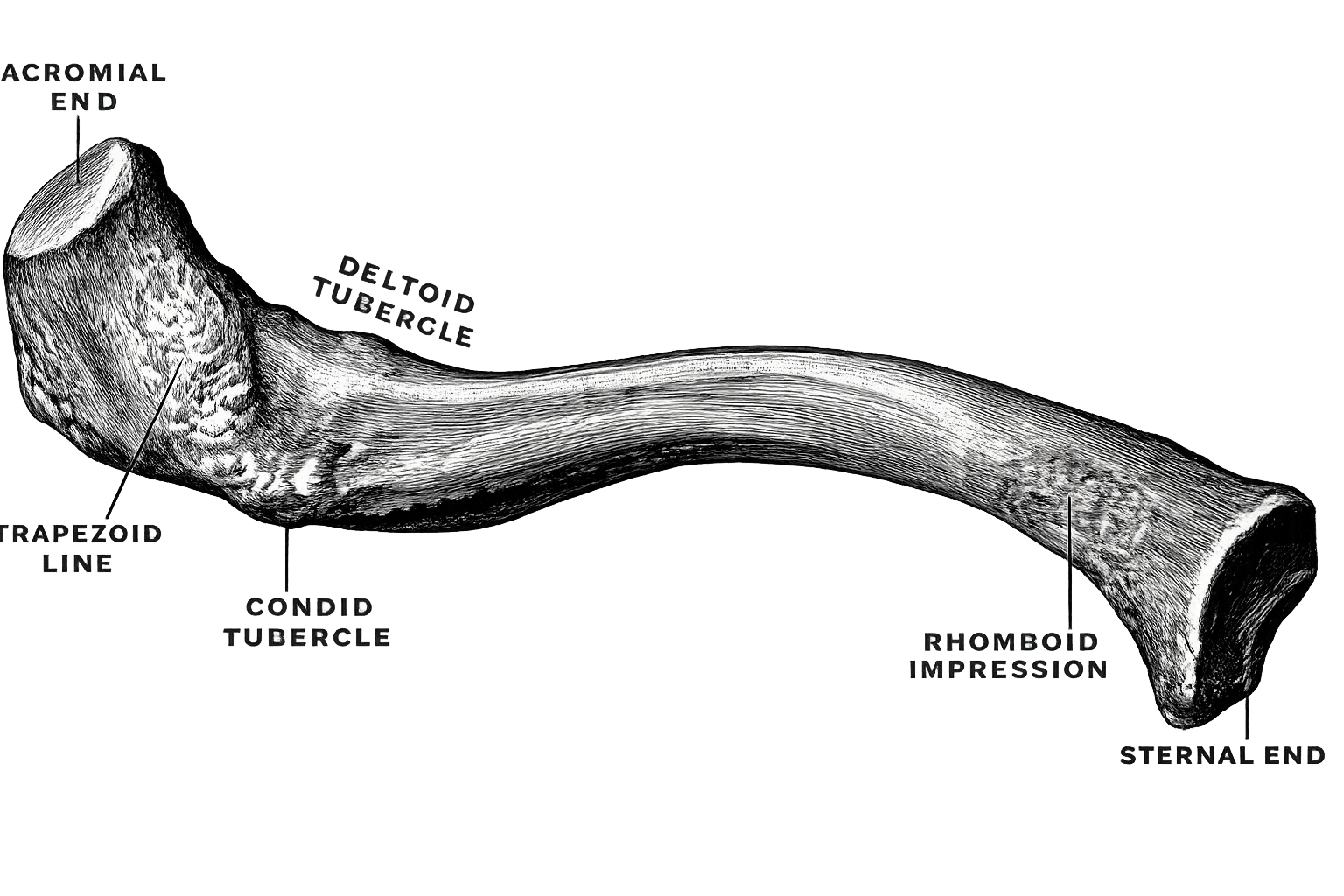
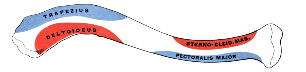
The Clavicle acts as an S-shaped strut between the upper limb and the axial skeleton, articulating medially with the sternum at the sternoclavicular joint and laterally with the acromion at the acromioclavicular joint [1][19]. It also provides attachment for several important muscles of the shoulder girdle and neck [1]. Medially, the clavicular head of the sternocleidomastoid arises from the superior surface of the clavicle, while the clavicular head of the pectoralis major originates from its anterior surface [1]. Laterally, the clavicular fibers of the deltoid arise from the anterior aspect of the lateral clavicle, whereas the trapezius attaches along its posterior aspect [1]. These major muscular attachment sites are illustrated in (Figure 1.8). Its inferior surface contains several clinically important landmarks, including the conoid tubercle and trapezoid line, which provide attachment for the two components of the coracoclavicular ligament, as well as the costoclavicular impression and subclavian groove[18]. These landmarks can be appreciated in the inferior view of the clavicle shown below (Figure 1.7). They are particularly relevant when considering clavicular fractures and surgical approaches, especially because the subclavian vessels and brachial plexus lie immediately deep to the clavicle [1][20].
Figure 1.7 — Inferior Surface of the Right Clavicle and Its Major Bony Landmarks
The inferior surface of the right clavicle showing the subclavian groove, conoid tubercle, trapezoid line, costoclavicular impression, and the articular surfaces of the sternal and acromial ends.
Source: Adapted from Frederick Henry Gerrish, Gerrish’s Text-book of Anatomy, Fig. 158, p. 134, 1902. Public-domain image obtained via Wikimedia Commons. Modification: the original background was removed for visual clarity; the anatomical illustration and labels were not otherwise altered.
Figure 1.8 — Muscular Attachments on the Superior Surface of the Right Clavicle
The illustration shows the principal areas of muscle attachment along the superior surface of the clavicle.
Adapted from Frederick Henry Gerrish, Gerrish’s Text-book of Anatomy, Fig. 157, p. 134, 1902, via Wikimedia Commons. Public-domain image. Modification: the original background was removed for visual clarity; the anatomical illustration and labels were not otherwise altered. Because the image is in the public domain, no Creative Commons licence is required. Attribution is still appropriate for academic and ethical transparency.
Proximal Humerus: Anatomy and Clinical Relevance
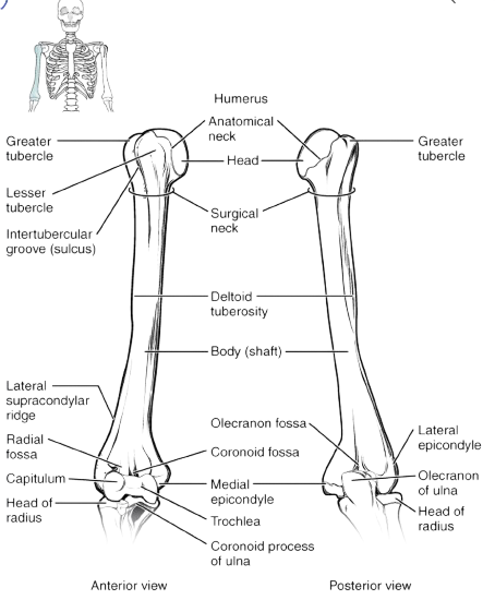
The humerus forms the bony foundation of the arm, and its proximal end forms the humeral side of the glenohumeral joint [1][9]. The rounded humeral head articulates with the glenoid cavity, while the anatomical neck marks the margin of the articular surface [1]. Immediately adjacent to it are the greater tubercle laterally and the lesser tubercle anteriorly [9]. These tubercles are particularly important in shoulder anatomy because they provide the bony attachment sites for the rotator cuff tendons: the supraspinatus, infraspinatus, and teres minor attach to the greater tubercle, whereas the subscapularis attaches to the lesser tubercle [1][11]. Between the two tubercles lies the intertubercular groove, or bicipital groove, which provides a passage for the tendon of the long head of the biceps [1][4]. Distal to the tubercles, the proximal humerus narrows at the surgical neck before continuing into the shaft [9]. These major landmarks are shown in (Figure 1.9).
These landmarks are not simply descriptive anatomy; they repeatedly become important when evaluating shoulder pathology [1]. The tubercles are directly related to rotator cuff function and may be involved in proximal humeral fractures [9], while the intertubercular groove is closely related to disorders of the long head of the biceps [4]. The surgical neck is also a clinically important region in proximal humeral fractures, where displacement may be associated with injury to nearby neurovascular structures, particularly the axillary nerve [9][10].
Figure 1.9 — Major Anatomical Landmarks of the Humerus and Elbow Joint
An anatomical overview of the humerus showing the humeral head, anatomical and surgical necks, greater and lesser tubercles, intertubercular groove, shaft, and distal humeral landmarks.
Adapted from OpenStax College, Anatomy & Physiology, 2013. Licensed under the Creative Commons Attribution 3.0 Unported licence (CC BY 3.0). Background removed by the author. Original source: Connexions/OpenStax. Licence: CC BY 3.0.
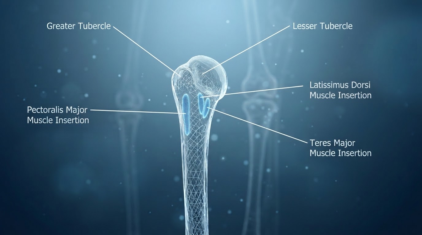
The region around the intertubercular groove is also an important site of muscular attachment [1]. The pectoralis major inserts along the lateral lip of the intertubercular groove, the latissimus dorsi attaches within the floor of the groove, and the teres major inserts along its medial lip [10]. Their close relationship to the proximal humerus is useful when interpreting tendon injuries and when identifying surgical anatomy around the anterior humerus [10]. These insertion sites are illustrated in (Figure 1.10).
Figure 1.10 — Crests of the Greater and Lesser Tubercles of the Humerus and the Insertion Sites of the Pectoralis Major, Teres Major, and Latissimus Dorsi Muscles
The illustration highlights the muscular attachment region surrounding the intertubercular groove of the proximal humerus.
Adapted from “Crests of Humerus” by Doctor Jana, 2019, via Wikimedia Commons. Original source: Doctor Jana — Humerus 3D Anatomy. Licensed under the Creative Commons Attribution–ShareAlike 4.0 International licence (CC BY-SA 4.0): CC BY-SA 4.0. Modification: the original background was removed for visual clarity. This adapted image is distributed under the same CC BY-SA 4.0 licence.
Rotator Cuff: Functional Anatomy, Innervation and Vascular Supply
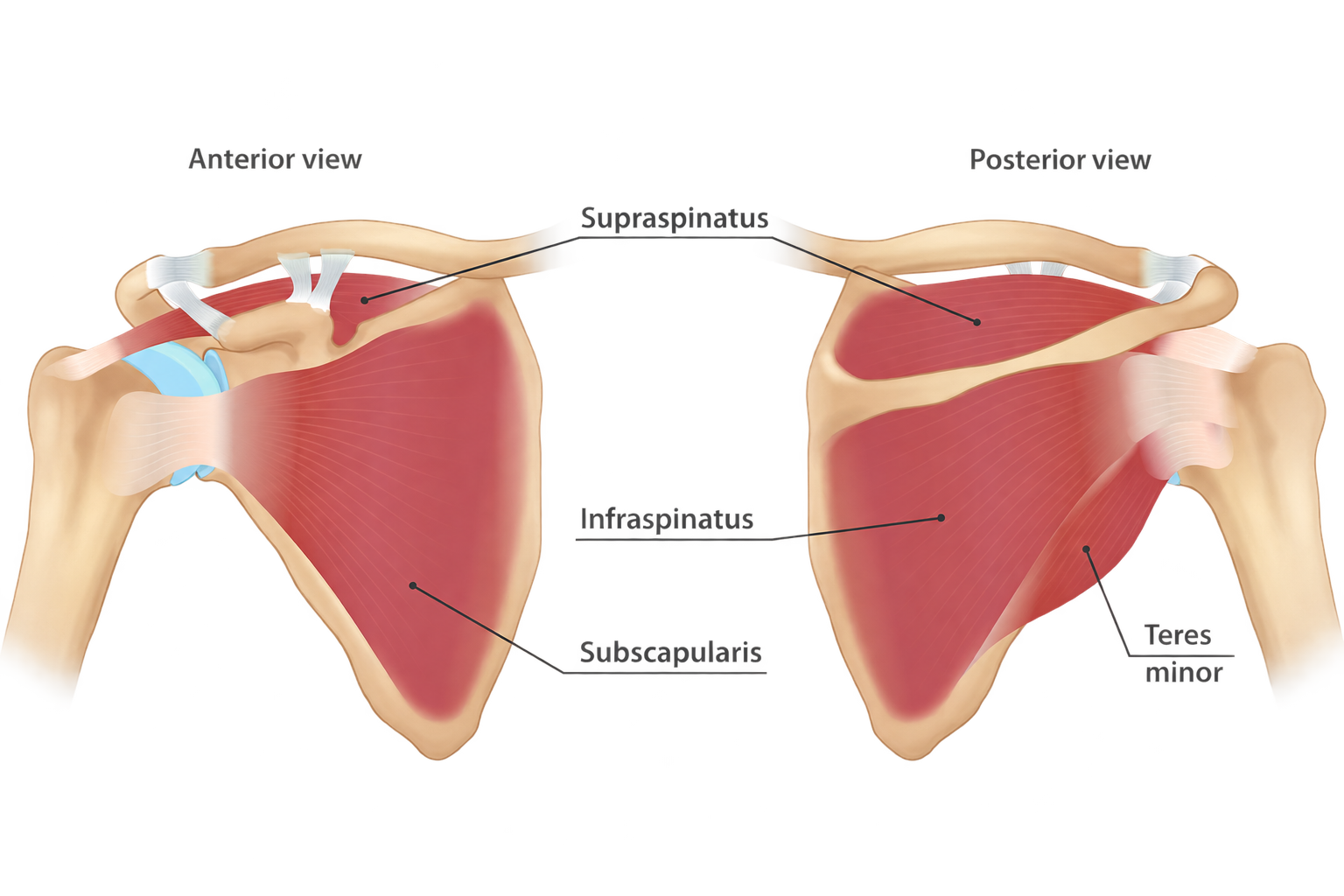
The rotator cuff is formed by the supraspinatus, infraspinatus, teres minor, and subscapularis. Their tendons blend around the humeral head and attach to the greater and lesser tubercles, creating a functional cuff around the glenohumeral joint. Beyond producing rotation and elevation, the cuff helps keep the humeral head centered on the glenoid while the larger superficial muscles move the arm. The principal muscle-tendon relationships are illustrated in (Figure 1.11)[11].
Supraspinatus
Attachment: Arises from the supraspinous fossa and inserts on the superior facet of the greater tubercle.
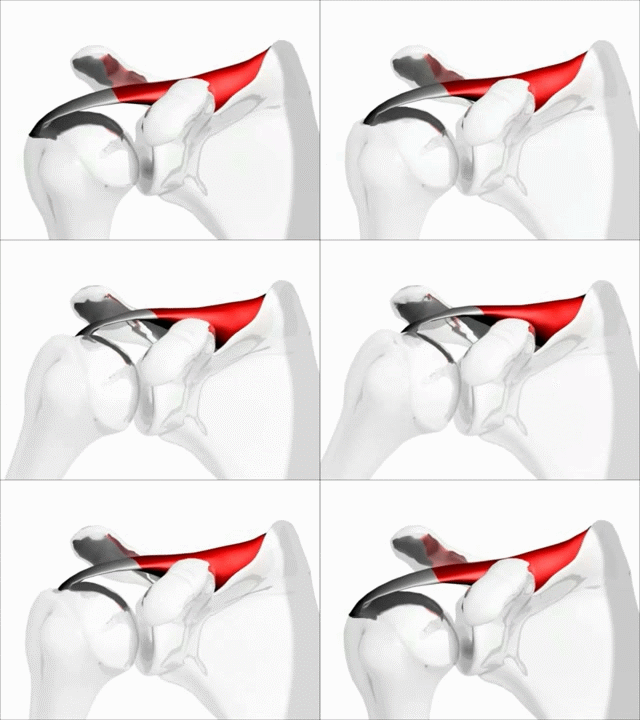
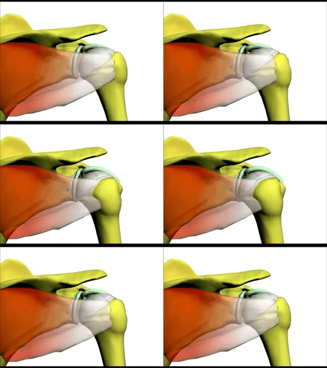
Role: Assists the initiation of shoulder abduction and continues to contribute during elevation, while also helping compress the humeral head against the glenoid. Its contribution to abduction is demonstrated in (Animation 1.12).
Arterial supply: Mainly through the suprascapular circulation, with an important contribution from the anterior circumflex humeral system to the tendon [13].
Clinical relevance: The supraspinatus is frequently involved in rotator cuff disease. Classic vascular studies describe relatively reduced vascularity near the distal tendon insertion, a region historically discussed in relation to degenerative cuff tearing [14].
Infraspinatus
Attachment: Originates from the infraspinous fossa and inserts on the middle facet of the greater tubercle.
Role: One of the principal external rotators of the humerus and an important posterior stabilizer of the glenohumeral joint.
Arterial supply: Mainly from the suprascapular artery, with contribution from the circumflex scapular circulation and the wider scapular anastomotic network [13].
Clinical relevance: Loss of infraspinatus function reduces external-rotation strength. Because its motor supply reaches the muscle after the suprascapular nerve courses around the scapula, neuropathy near the spinoglenoid region may disproportionately affect infraspinatus function.
Teres Minor
Attachment: Arises from the lateral border of the scapula and inserts on the inferior facet of the greater tubercle.
Role: Contributes to external rotation and posterior stabilization of the humeral head, working closely with the infraspinatus.
Arterial supply: Supplied through the posterior shoulder vascular network, particularly branches associated with the posterior circumflex humeral and circumflex scapular arteries [13].
Clinical relevance: Teres minor function becomes especially important when the posterosuperior cuff is deficient. Its anatomy and external-rotation activity are relevant in massive cuff tears and reverse shoulder arthroplasty [16].
Subscapularis
Attachment: Originates from the subscapular fossa and inserts primarily on the lesser tubercle of the humerus.
Role: The principal internal rotator within the rotator cuff and a major contributor to anterior glenohumeral stability.
Innervation: Upper and lower subscapular nerves, predominantly C5–C6 [12].
Arterial supply: The subscapular artery is a major contributor, with additional supply from the suprascapular, circumflex scapular, lateral thoracic, and posterior circumflex humeral systems [15].
Clinical relevance: Tears involving the superior subscapularis may occur together with pathology of the bicipital pulley and long head of the biceps, so the anterior cuff deserves careful assessment when biceps instability is suspected [4].
Figure 1.11 — Anterior and Posterior Views of the Rotator Cuff Musculature
Anterior and posterior perspectives showing the supraspinatus, infraspinatus, teres minor, and subscapularis around the glenohumeral joint.
Modified (background removed) from the original by InjuryMap, via Wikimedia Commons. This modified image is licensed under the Creative Commons Attribution–ShareAlike 4.0 International licence (CC BY-SA 4.0): CC BY-SA 4.0.
Animation 1.12 — Action of the Supraspinatus Muscle in Shoulder Abduction
Animated demonstration of the supraspinatus contribution to shoulder abduction.
Moon YL. Shoulder motion with rotator cuff (supraspinatus). 2008. Uploaded to Wikimedia Commons / YouTube under the Creative Commons Attribution 3.0 Unported licence (CC BY 3.0). Available via Wikimedia Commons: Shoulder motion with rotator cuff (supraspinatus). Licence: CC BY 3.0.
Functional Myology and Force Couples
Around the humeral head, the rotator cuff tendons blend into a continuous musculotendinous covering that contributes to dynamic control of the joint [5]. Burkhart and colleagues used the “suspension bridge” concept to describe the biomechanical role of the rotator cable within this system [6].
Glenohumeral Joint Capsule, Glenohumeral Ligaments and Labrum
Glenoid Cavity of the Scapula
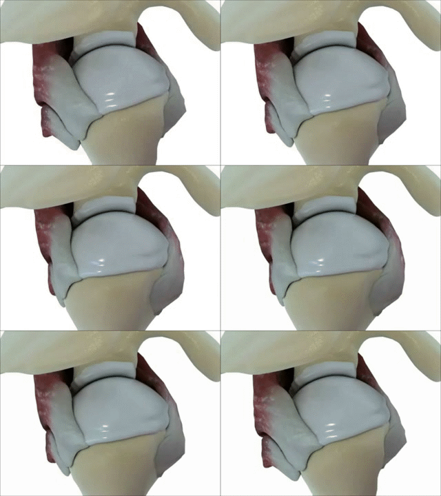
The glenoid cavity is a shallow, pear-shaped articular surface on the lateral aspect of the scapula. Its contour is generally narrower superiorly and broader inferiorly, creating the characteristic pear-shaped appearance of the glenoid [1]. The characteristic pear-shaped outline and location of the glenoid cavity can be appreciated in the animation below.
Functionally, the glenoid cavity forms the socket of the glenohumeral joint by articulating with the humeral head. Its relatively shallow geometry permits the extensive range of motion that characterizes the shoulder, but it provides little stability through bony shape alone. Glenohumeral stability therefore also depends on the glenoid labrum, joint capsule, glenohumeral ligaments, and the coordinated action of the surrounding musculature [1].
Figure 1.12A — Glenoid Cavity of the Left Scapula
Animated three-dimensional representation of the left scapula highlighting the glenoid cavity.
Source: BodyParts3D, Database Center for Life Science (DBCLS). Original file:
Glenoid cavity of left scapula - animation.gif.
Created 3 May 2013. Licensed under the
Creative Commons Attribution-ShareAlike 2.1 Japan licence (CC BY-SA 2.1 JP).
No changes were made to the original GIF. Any reuse or redistribution of this image must comply with the attribution and ShareAlike terms of the licence. Attribution does not imply endorsement by BodyParts3D or DBCLS.
The glenohumeral joint depends on a coordinated group of static stabilizers that complement the dynamic action of the rotator cuff. The capsule forms a relatively compliant fibrous envelope around the joint, while localized capsular thickenings form the superior, middle, and inferior glenohumeral ligaments. Around the glenoid rim, the labrum provides a fibrocartilaginous interface between the bony socket and the capsuloligamentous structures [21][23].
The joint capsule surrounds the humeral head and glenoid and is deliberately lax enough to permit the shoulder's large range of motion. Medially, it is attached around the glenoid rim and blends with the labral-capsular complex; laterally, it attaches around the proximal humerus near the anatomical neck. Its inferior portion is more redundant and forms the axillary pouch, which unfolds as the arm elevates [1][22].
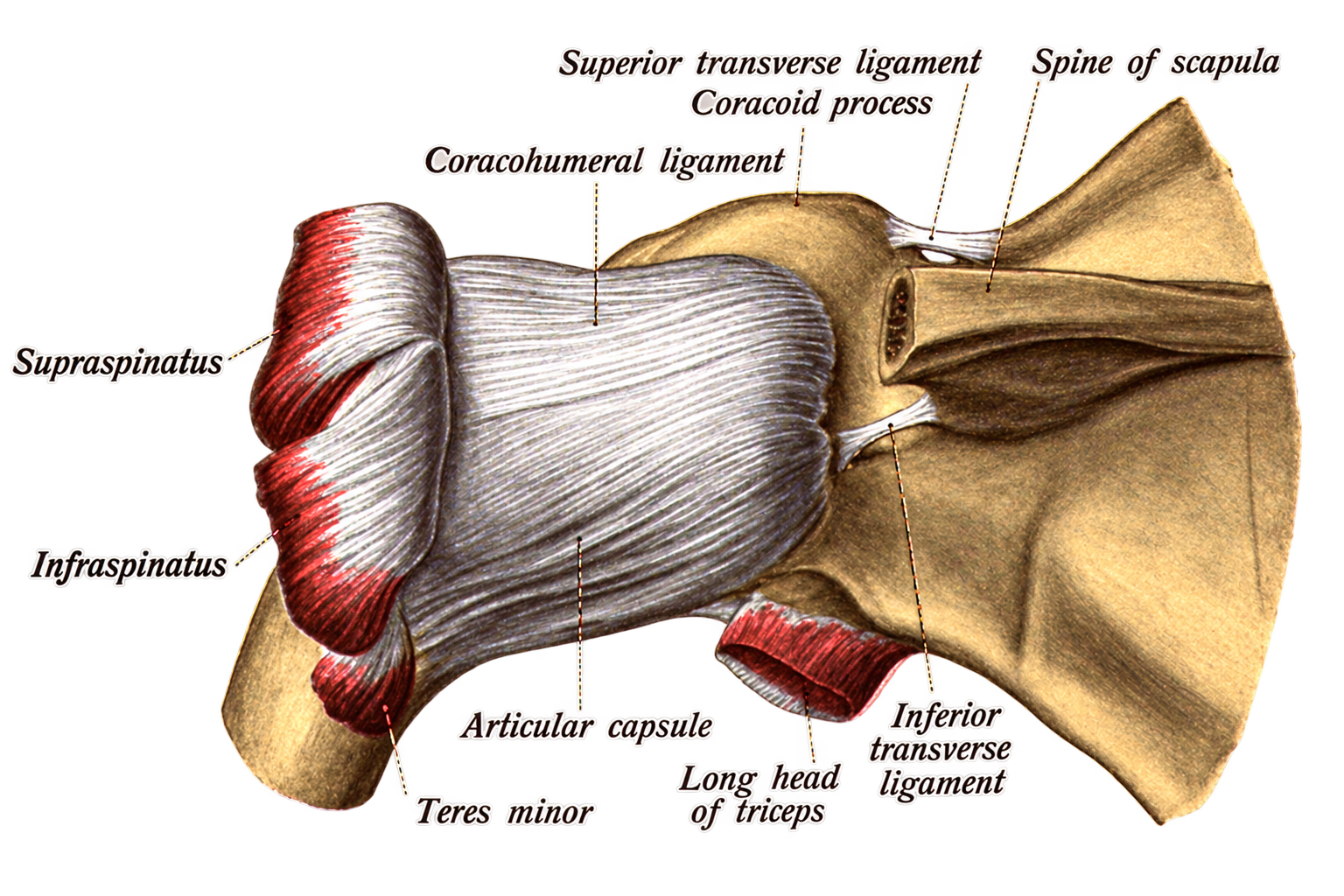
Functionally, the capsule is not equally tight in every position. Different regions become tensioned as the humerus moves through abduction and rotation, allowing mobility in one position while providing restraint in another [21]. The articular joint capsule and its surrounding anatomical relationships are illustrated in Figure 1.13A.
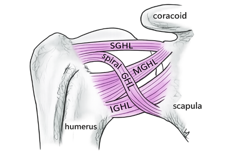
The superior (SGHL), middle (MGHL), and inferior glenohumeral ligaments (IGHL) are specialized thickenings of the capsule. Their relative positions are shown in Figure 1.13. Their stabilizing role changes with arm position rather than functioning as three identical restraints [21].
Superior Glenohumeral Ligament
Most relevant when the arm is near the side. It contributes to restraint of inferior translation and excessive external rotation in lower degrees of abduction.
Middle Glenohumeral Ligament
Located across the anterior capsule and shows considerable anatomic variation. It contributes to anterior stability particularly through the mid-range of abduction.
Inferior Glenohumeral Ligament Complex
Composed of an anterior band, posterior band, and intervening axillary pouch. It becomes especially important as the arm is abducted; the anterior band is a major restraint to anterior translation in abduction and external rotation [22].
Clinical relevance: Because the inferior complex is a major stabilizer in the abducted shoulder, injury to the anteroinferior capsulolabral region is central to traumatic anterior instability and is an important structure to recognize during arthroscopic stabilization.
The glenoid labrum is a fibrocartilaginous rim surrounding the glenoid. It enlarges the effective articular margin and provides an attachment region for the capsule and glenohumeral ligaments. The superior labrum is closely related to the origin of the long head of the biceps, whereas the inferior labrum is more firmly attached to the glenoid rim [23].
This anatomy helps explain why injury at different portions of the labrum can produce different clinical patterns: the superior labrum is closely linked to the biceps anchor, while the anteroinferior labrum works with the inferior glenohumeral ligament complex in resisting instability.
Figure 1.13 — Glenohumeral Capsule, Labrum and Glenohumeral Ligaments
The image demonstrates the relative location of the superior, middle, and inferior glenohumeral ligaments around the capsule and their relationship to the glenoid labrum.
Local educational image: gleno.jpg. The image is referenced from the text to identify the location of the glenohumeral ligaments. Source and licence metadata were not supplied with the image and should be added before final publication.
Figure 1.13A — Articular Joint Capsule and Its Surroundings (posterior view)
Anatomical illustration of the shoulder joint capsule and its relationship to the surrounding structures.
Illustration title:
Anatomical illustration of the shoulder joint from Sobotta's Atlas and Text-book of Human Anatomy (1909 edition).
Citation:
Sobotta J. (1909). Atlas and Text-book of Human Anatomy (English terminology ed.). Public Domain archive scan hosted on Wikimedia Commons.
Other Clinically Important Ligaments of the Shoulder
Several additional ligamentous structures become important when interpreting acromioclavicular injury, subacromial anatomy, and pathology of the long head of the biceps. Their importance is less about acting as one single stabilizing unit and more about how each structure supports a particular region of the shoulder.
Acromioclavicular Ligament
The acromioclavicular (AC) ligament reinforces the capsule of the AC joint and contributes to local stability between the lateral clavicle and acromion. The superior component is particularly robust, while the capsuloligamentous complex as a whole contributes to control of horizontal translation at the AC joint [3].
In traumatic AC separation, the AC capsuloligamentous complex is injured early, while increasing injury severity may extend to the coracoclavicular ligaments.
Conoid Ligament
The conoid ligament is the more medial component of the coracoclavicular ligament complex. It extends from the coracoid toward the conoid tubercle of the clavicle and is an important part of the suspensory linkage between scapula and clavicle [18].
Its position is clinically important when reconstructing the coracoclavicular ligaments after higher-grade AC joint injuries.
Trapezoid Ligament
The trapezoid ligament forms the more lateral component of the coracoclavicular complex, attaching between the coracoid and the trapezoid line of the clavicle [18].
Together with the conoid ligament, it helps preserve the normal relationship between the clavicle and scapula and provides key anatomic landmarks during AC joint reconstruction.
Coracoacromial Ligament
The coracoacromial ligament spans from the coracoid process to the acromion and forms the soft-tissue component of the coracoacromial arch above the humeral head [18].
Its relationship to the subacromial space makes it clinically relevant in rotator cuff disease and in procedures involving the anterior acromion.
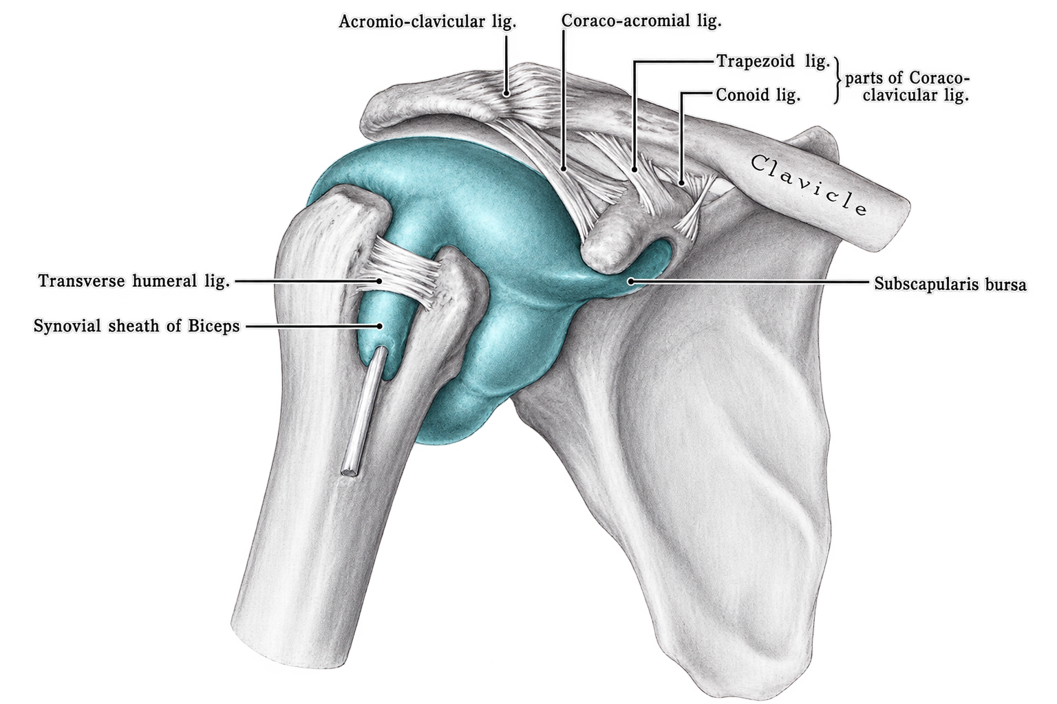
The positions of the AC, coracoclavicular, and coracoacromial ligamentous structures can be reviewed in Figure 1.14.
Figure 1.14 — Clinically Important Ligaments of the Shoulder
Image used to orient the reader to the acromioclavicular ligament, the conoid and trapezoid components of the coracoclavicular ligament complex, the coracoacromial ligament, and the structural relationship between the transverse humeral ligament and the long head of the biceps tendon within the bicipital groove.
Source: Uploaded by AHMED MAHER BAKRI via Wikimedia Commons. Licensed under the Creative Commons Attribution–ShareAlike 4.0 International licence (CC BY-SA 4.0). Licence: CC BY-SA 4.0.
Transverse Humeral Ligament and the Long Head of the Biceps
Traditionally, the transverse humeral ligament (THL) is described as a band crossing the intertubercular groove and forming a roof over the long head of the biceps tendon. This description explains its close clinical association with the bicipital groove and with disorders in which the biceps tendon becomes unstable or displaced. The structural relationship between the transverse humeral ligament and the long head of the biceps tendon within the bicipital groove can be viewed in Figure 1.14.
Modern anatomic studies, however, have questioned whether the THL is consistently present as a completely separate ligament. Cadaveric investigations have shown that the tissue crossing the groove can include extensions of the subscapularis and supraspinatus tendons as well as fibrous expansions associated with the pectoralis major [24][25].
For that reason, stability of the long head of the biceps is better understood as the result of a bicipital pulley complex rather than the transverse humeral ligament acting alone. The superior glenohumeral ligament, coracohumeral ligament, and adjacent fibers of the subscapularis and supraspinatus contribute to this stabilizing system; disruption can permit abnormal medial translation, subluxation, or dislocation of the biceps tendon [4].
Clinical relevance: When biceps instability is suspected, the surrounding pulley and subscapularis should be assessed rather than considering the transverse humeral ligament in isolation.
Periarticular Bursae of the Shoulder: Anatomy and Clinical Relevance
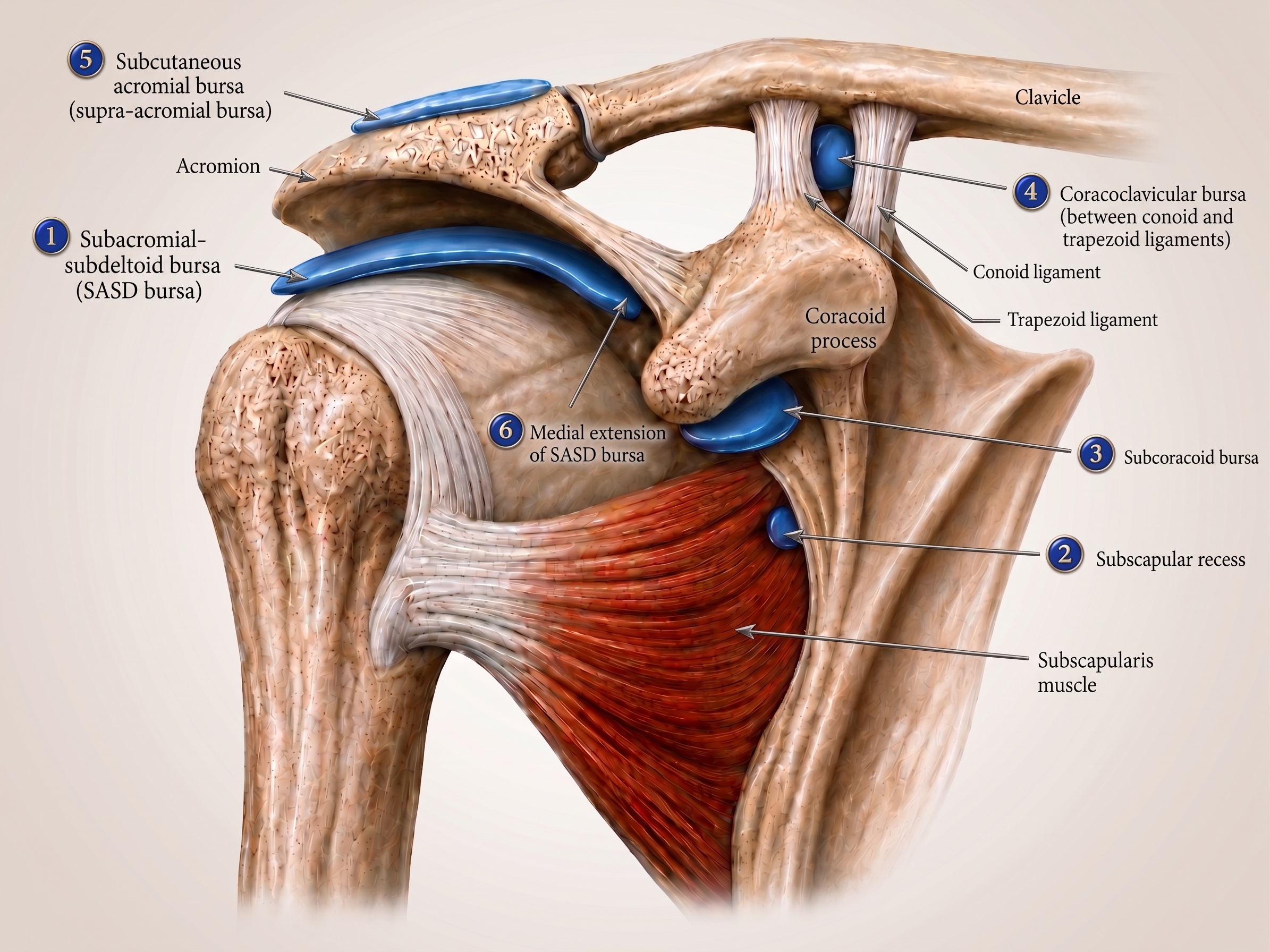
The shoulder contains several synovial-lined bursae and recesses positioned at interfaces where tendons, muscles, bone, and ligamentous structures move against one another. Their principal mechanical role is to reduce friction and permit smooth tissue gliding during the shoulder's large range of motion. The clinically most important structures include the subacromial-subdeltoid (SASD) bursa, subscapular recess, subcoracoid bursa, coracoclavicular bursa, and supra-acromial bursa[40].
Subacromial-Subdeltoid Bursa (SASD)
The subacromial-subdeltoid bursa is the largest and most clinically important bursa around the shoulder. It lies superficial to the rotator cuff, particularly the supraspinatus tendon, and deep to the acromion, coracoacromial arch, acromioclavicular region, and deltoid. Its subacromial and subdeltoid portions form a broad gliding plane that reduces friction as the greater tuberosity and rotator cuff pass beneath the coracoacromial arch during elevation of the arm [40].
In the normal shoulder, the SASD bursa does not communicate with the glenohumeral joint. Distension or inflammatory change may occur with subacromial bursitis, rotator cuff disease, mechanical subacromial impingement, infection, trauma, or reactive inflammatory processes. In a full-thickness rotator cuff tear, disruption of the intervening cuff can establish communication between the glenohumeral joint and the SASD bursa, allowing joint fluid to enter the bursal space [40].
Subscapular Recess
The subscapular recess, often termed the superior subscapularis recess, is a synovial extension of the glenohumeral joint related to the superior portion of the subscapularis. This distinction is important because the recess normally communicates with the joint cavity and should not be confused on imaging with the adjacent subcoracoid bursa, which ordinarily does not communicate with the glenohumeral joint [41].
Fluid within the recess may therefore reflect glenohumeral joint fluid rather than primary bursitis. Marked fluid distension of the superior subscapularis recess has also been described as an MRI sign associated with adhesive capsulitis, particularly during the painful freezing phase, although it should be interpreted together with the other clinical and imaging features of frozen shoulder [42].
Subcoracoid Bursa
The subcoracoid bursa occupies the anterior shoulder beneath the coracoid process and is closely related to the anterior surface of the subscapularis. Unlike the subscapular recess, it does not normally communicate with the glenohumeral joint, although an anatomic communication with the SASD bursa may occasionally occur. Its position allows it to reduce friction around the subscapularis and adjacent coracoid structures during shoulder motion [40][43].
Pathologic distension of the subcoracoid bursa may represent subcoracoid bursitis and can accompany anterior shoulder pathology. MRI studies have demonstrated an association between subcoracoid effusion and tears of the anterior rotator cuff, including lesions involving the rotator interval. Correctly distinguishing this bursa from the communicating subscapular recess is therefore important when interpreting fluid in the subcoracoid region [44].
Coracoclavicular and Supra-acromial Bursae
The coracoclavicular bursa and supra-acromial bursa are smaller and less frequently encountered than the SASD bursa. The coracoclavicular bursa lies in the coracoclavicular region, while the supra-acromial bursa is superficial to the acromion. Both are recognized components of the normal periarticular bursal anatomy of the shoulder [40]. Because these bursae are small and are not routinely conspicuous on imaging, their pathologic distension is much less commonly encountered; when enlarged, a focal bursal process such as inflammation, repetitive mechanical irritation, or trauma should be considered in the appropriate clinical context rather than assuming that all superior shoulder fluid represents SASD bursitis.
Clinical perspective: Bursal fluid should be interpreted according to its precise anatomic compartment. SASD fluid may accompany bursitis, impingement, or rotator cuff tearing; subcoracoid fluid may point toward anterior cuff or rotator-interval disease; and fluid in the subscapular recess can simply track from the glenohumeral joint or become conspicuous in conditions such as adhesive capsulitis [40][42][44].
Figure 1.15 — Bursae and Ligamentous Anatomy of the Anterior Right Shoulder.
The blue regions represent the different bursae and synovial recesses surrounding the shoulder joint. Note: Generated by author using Biorender.
Shoulder Joint Articulations
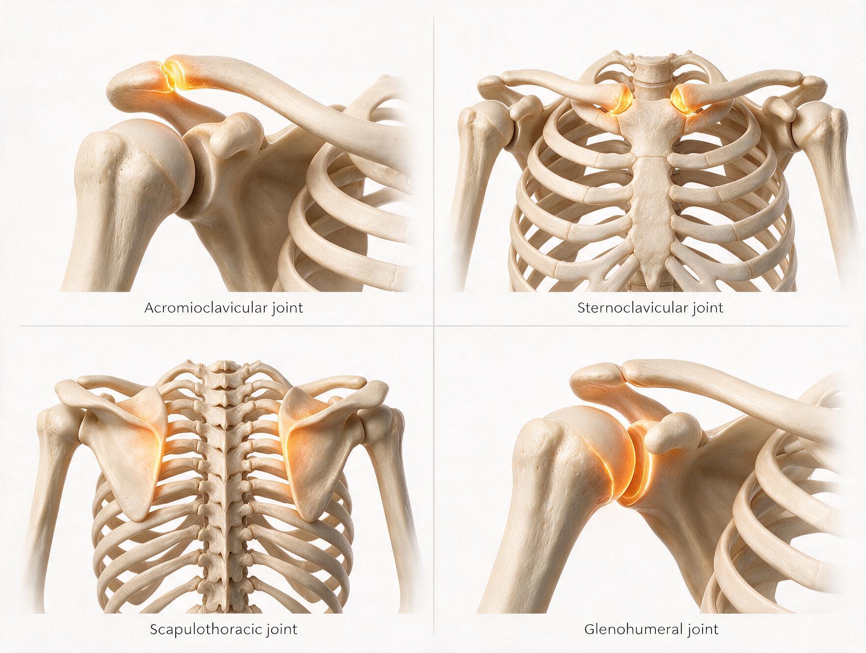
The shoulder should be understood as a four-articulation functional complex, not as the glenohumeral joint alone. The sternoclavicular (SC), acromioclavicular (AC), scapulothoracic (ST), and glenohumeral (GH) articulations coordinate clavicular, scapular, and humeral motion so that the hand can be positioned through a very large three-dimensional workspace. Three are true synovial joints (SC, AC, and GH); the scapulothoracic articulation is a physiologic sliding interface rather than a true synovial joint [1][54].
Acromioclavicular Joint
Synovial plane joint
The acromioclavicular joint is formed by the lateral end of the clavicle and the medial acromion. Its key functional role is to allow the scapula to adjust relative to the clavicle as the arm moves. Although its articular excursion is small compared with the glenohumeral joint, these fine rotational and translational adjustments are mechanically essential during arm elevation. The AC capsule and acromioclavicular ligaments contribute importantly to horizontal stability, whereas the coracoclavicular ligaments—the conoid and trapezoid—are major restraints to vertical displacement of the scapula–clavicle complex [48].
Primary role
Transfers load between scapula and clavicle while allowing the scapula to upwardly rotate, tilt, and internally/externally rotate during elevation [48].
Key stabilizers
AC capsuloligamentous complex for horizontal control; conoid and trapezoid portions of the coracoclavicular ligament complex for vertical stability [48].
Clinical localization
Symptomatic disease typically produces focal superior shoulder pain and tenderness, often reproduced by cross-body adduction.
Major disease and injury patterns
AC separation / instability: traumatic disruption ranges from ligament sprain to complete displacement; the Rockwood system is commonly used to describe injury severity [48].
AC osteoarthritis: degeneration is common and may be symptomatic or incidental; clinical correlation is therefore essential [49].
Distal clavicle osteolysis: a characteristic overuse/stress-related disorder, especially in weight-training athletes exposed to repeated AC loading [50].
Clinical pearl: Radiographic AC degeneration is not automatically the pain generator. The history, focal examination, provocative testing, imaging, and—when needed—a selective diagnostic injection should agree before symptoms are attributed to the AC joint [49].
Sternoclavicular Joint
Synovial saddle joint
The sternoclavicular joint joins the medial clavicle to the manubrium and first costal cartilage. Functionally, it links the upper limb to the axial skeleton and is the only true osseous connection between them. An intra-articular fibrocartilaginous disc improves congruity and separates the joint space, while the sternoclavicular, interclavicular, and costoclavicular ligaments provide substantial restraint [51].
Motion
Allows clavicular elevation/depression, protraction/retraction, and axial rotation; these motions are required for normal scapular positioning during arm elevation [51].
Stability concept
Despite limited articular congruity, the joint is strongly stabilized by its capsule, disc, and ligament complex; the posterior capsular structures are particularly important in resisting displacement [52].
Why it matters
Loss of normal SC motion can disturb the entire shoulder girdle because clavicular motion is mechanically linked to scapular motion [51].
Major disease and injury patterns
Traumatic instability and dislocation: anterior displacement is more common, but posterior dislocation is clinically more dangerous because of the adjacent mediastinal neurovascular, airway, and visceral structures [52].
Osteoarthritis: degenerative change may cause localized pain, swelling, crepitus, and pain with shoulder-girdle motion [51].
Septic arthritis: uncommon but potentially serious because infection can extend into adjacent chest-wall and mediastinal structures; Staphylococcus aureus is the dominant pathogen in reported series [53].
Atraumatic inflammatory disorders: the SC joint may also be involved in inflammatory arthropathies and SAPHO-spectrum disease [51].
Safety pearl: A suspected posterior SC dislocation deserves urgent recognition. CT is substantially more useful than routine radiography for defining displacement and its relationship to mediastinal structures [52].
Scapulothoracic Articulation
Functional articulation
The scapulothoracic articulation is not a true synovial joint: there is no opposing cartilage-lined joint surface, capsule, or conventional synovial cavity between the scapula and thorax. Instead, the scapula glides over the posterolateral rib cage through organized muscular and bursal planes, particularly the interface between subscapularis and serratus anterior. Its central functional role is to position and rotate the scapula on the thorax, continually reorienting the glenoid so that glenohumeral motion remains efficient [54].
Scapular motions
Upward/downward rotation, internal/external rotation, anterior/posterior tilt, elevation/depression, and protraction/retraction combine during functional arm motion [55].
Muscular control
Serratus anterior and trapezius are central to upward rotation and stable scapular positioning, with additional contributions from the rhomboids, levator scapulae, and pectoralis minor [54].
Scapulohumeral rhythm
Arm elevation reflects coordinated GH and scapular motion. The classic approximately 2:1 GH-to-scapular description is a useful teaching model, but actual ratios vary across the arc and between individuals [5][55].
Major disease and dysfunction patterns
Scapular dyskinesis: an alteration in scapular position or motion that can accompany rotator cuff disease, impingement-related pain, labral pathology, instability, and other shoulder disorders; it should be viewed as an impairment pattern rather than a single disease diagnosis [56].
Scapular winging: may result from dysfunction of key periscapular muscles or their nerves, classically serratus anterior/long thoracic nerve or trapezius/spinal accessory nerve dysfunction [54].
Snapping scapula / scapulothoracic bursitis: painful crepitus may arise from inflamed bursae, repetitive overuse, altered scapular mechanics, or osseous/soft-tissue abnormalities along the scapulothoracic interface [57].
Clinical pearl: The scapula is the moving base of the glenoid. When its position or motion changes, apparent “glenohumeral” symptoms may partly reflect altered scapular mechanics; therefore observation of scapular motion should be part of a complete shoulder examination [56].
Glenohumeral Joint
Synovial ball-and-socket joint
The glenohumeral joint is the principal articulation between the humeral head and glenoid and provides the largest component of humeral mobility within the shoulder complex. It permits exceptional multiaxial motion but has relatively little intrinsic bony constraint because the humeral head is large and the glenoid is shallow. Stability is therefore shared between static structures—labrum, capsule, glenohumeral ligaments, joint geometry and negative intra-articular pressure—and dynamic muscular control, particularly the rotator cuff [1][26].
Primary role
Produces the largest component of shoulder flexion, extension, abduction/adduction, and internal/external rotation while the scapula simultaneously repositions the glenoid.
Dynamic centering
Rotator-cuff contraction creates compressive and balancing forces that help keep the humeral head centered during motion [26][28].
Mobility–stability trade-off
The same anatomy that permits enormous motion also makes the GH joint particularly dependent on intact soft-tissue stabilizers and coordinated neuromuscular control.
Major disease and injury patterns
Instability and dislocation: failure of normal humeral-head centering may follow traumatic capsulolabral injury and may be compounded by glenoid or humeral-head bone loss; recurrent instability can produce progressive soft-tissue and osseous damage [60].
Glenohumeral osteoarthritis: progressive articular degeneration can produce pain, stiffness, joint-space narrowing, osteophytes, and characteristic glenoid/humeral remodeling [58].
Adhesive capsulitis (frozen shoulder): a painful restriction of both active and passive motion related to capsular contracture; it may be primary or secondary [59].
Inflammatory, infectious, and osteonecrotic processes may also damage the GH articular surfaces and accelerate secondary degeneration [58].
Biomechanical pearl: The GH joint cannot be interpreted independently of the scapula. Humeral motion changes the load on the cuff and capsule, while scapular position changes glenoid orientation; normal shoulder function therefore depends on coordinated GH–ST motion rather than isolated ball-and-socket movement [55].
Figure 1.16 — Four Major Articulations of the Shoulder
Note: Generated by author using BioRender.
Rotator Cuff Biomechanics
Concept 1 — Concavity–Compression and Dynamic Humeral Head Centering
Concavity–compression is a central mechanism of dynamic glenohumeral stability. Because the rounded humeral head articulates with a relatively shallow glenoid, stability depends not only on the geometry of the joint but also on compressive forces that keep the articular surfaces opposed
[26][27].
In the classic quantitative model of concavity–compression, increasing compression of the humeral head into the glenoid concavity increases the tangential force required to translate the head across the glenoid rim
[26].
Experimental work further demonstrated that the glenoid labrum deepens the effective concavity and contributes measurably to the stabilizing effect of concavity–compression
[27].
The rotator cuff provides an important muscular source of this dynamic compression. When the supraspinatus, infraspinatus, teres minor, and subscapularis contract, their combined force components compress the humeral head toward the glenoid and resist unwanted translation while motion continues
[28][26].
Cadaveric analysis has shown that the individual cuff muscles make substantial contributions to dynamic stability in both the mid-range and end-range of motion, with their relative importance changing according to joint position
[28].
Functionally, this coordinated compression helps maintain humeral-head centering while preserving the large rotational and elevational excursions required of the shoulder
[26][28].
The concavity–compression mechanism is demonstrated in Figure 1.17.
Figure 1.17 — Concavity Compression Mechanism
Interactive 3D demonstration of rotator cuff–generated concavity compression. Use the compressive-force control to observe progressive cuff contraction, humeral-head centring, and dynamic stabilization against the glenoid.
Interactive educational 3D module integrated into this e-book. Drag to rotate, scroll to zoom, and use the controls to show or hide labels and force vectors. For a historical animation of the same biomechanical mechanism, see Moon YL. Concavity compression of shoulder joint; working as a dynamic stabilizer [Video/Animation]. 2016
[29].
Concept 2 — Coronal Plane Rotator Cuff Force Couple
During arm elevation, the glenohumeral joint must generate enough torque to raise the humerus while simultaneously limiting excessive superior translation of the humeral head
[5][31].
In the coronal plane force-couple model, the superiorly directed action of the deltoid is balanced by the inferiorly directed stabilizing components of the rotator cuff, particularly the infraspinatus, teres minor, and subscapularis[30][31].
The resulting balance allows the deltoid and cuff to contribute to elevation while maintaining a stable glenohumeral fulcrum
[28][31].
The deltoid is a major motor of arm elevation, but its line of pull also produces a superior translational tendency at the humeral head
[31].
The inferior cuff supplies a counteracting component that limits superior migration and helps preserve humeral-head centering throughout elevation
[28][31].
In a cadaveric shoulder model, simulated abduction with the deltoid alone—or without the stabilizing contribution of the infraspinatus, teres minor, and subscapularis—produced significantly greater superior displacement of the humeral head than conditions in which the cuff was functionally intact
[31].
The interaction between the superiorly directed deltoid vector and the counterbalancing inferior cuff force is illustrated in Figure 1.18[32].
Biomechanical summary:
Deltoid-driven elevation creates a superior translational tendency, whereas the inferior rotator cuff supplies a counterbalancing stabilizing component. Their coordinated action maintains a functional center of rotation and permits efficient arm elevation without excessive superior humeral-head migration.
Figure 1.18 — Coronal Plane Rotator Cuff Force Couple
As the deltoid generates a superior translation vector, the inferior rotator cuff (infraspinatus, teres minor) creates a counteracting inferior force to maintain the humeral head centered within the glenoid.
Note: generated by author using BioRender.
Concept 3 — Rotator Cable–Crescent Complex
The rotator cable–crescent complex describes a distinctive structural organization within the posterosuperior rotator cuff. Burkhart, Esch, and Jolson described the rotator crescent as the relatively thin, crescent-shaped distal portion of the supraspinatus and infraspinatus tendons and the rotator cable as a thicker band of fibers forming the proximal margin of this crescent
[6].
In their cadaveric description, the cable was substantially thicker than the adjacent crescent and was proposed to function as a load-transmitting structure across the cuff
[6].
This arrangement led to the influential “suspension bridge” model, in which the rotator cable was proposed to transfer tensile load between its anterior and posterior attachments while partly stress-shielding the thinner crescent
[6].
Later cadaveric testing supported an important load-bearing role for the anterior cable insertion: tears involving the anterior cable produced greater tear gapping and altered regional tendon strain compared with equivalently sized crescent tears
[33].
The suspension-bridge interpretation should not, however, be presented as mechanically absolute. A more recent cadaveric study found that releasing the rotator cable did not measurably change cable or crescent strain during abduction and concluded that the crescent itself remained important for transmitting abduction force to the greater tuberosity
[34].
The cable–crescent concept therefore remains highly useful for describing cuff anatomy and tear location, while the exact degree to which the cable stress-shields the crescent remains an area of ongoing biomechanical investigation
[33][34].
Clinical relevance:
Describing whether a tear involves the cable, the crescent, or both can add biomechanical context beyond tear dimensions alone, particularly when assessing the anterior supraspinatus cable attachment and the functional continuity of the posterosuperior cuff.
Concept 4 — Transverse Force Couple
In the transverse plane, the anterior rotator cuff and posterior rotator cuff generate opposing rotational forces around the humeral head. The subscapularis forms the anterior component, while the infraspinatus and teres minor form the principal posterior component. Their balanced activity permits internal and external rotation while helping maintain the humeral head centered against the glenoid rather than allowing uncontrolled anterior or posterior translation
[35][36].
The functional importance of this balance has been demonstrated experimentally. In a cadaveric deficiency model, glenohumeral ball-and-socket kinematics were maintained when the transverse force couple—subscapularis anteriorly and infraspinatus/teres minor posteriorly—remained intact, whereas disruption of this force couple produced significant alterations in glenohumeral motion
[35].
Imaging-based studies provide complementary anatomic support: CT assessment found no significant difference between the muscle volumes of the anterior and posterior components in nonpathologic shoulders
[36],
while MRI measurements demonstrated significant correlations between the cross-sectional areas of the muscles forming the transverse force couple, supporting the concept of a dynamically balanced shoulder
[37].
The transverse force couple can be visualized from complementary perspectives. Figure 1.19 demonstrates this concept from a lateral view, showing the relationship between the anterior subscapularis and the posterior infraspinatus/teres minor around the humeral head
[38].
Figure 1.20 shows the transverse force-couple concept from a posterior view, emphasizing the posterior component formed by the infraspinatus and teres minor
[39].
Biomechanical summary:
The transverse force couple is not simply a contest between internal and external rotators. It is a coordinated anterior–posterior balance that allows rotation while preserving a stable glenohumeral fulcrum. Loss of either the anterior or posterior component can therefore disturb both rotational strength and joint centering.
Figure 1.19 — Transverse Force Couple of the Rotator Cuff (Lateral view)
Lateral view of the right glenohumeral joint demonstrating the anterior subscapularis and posterior infraspinatus and teres minor components of the transverse rotator-cuff force couple.
Moon, Y. L. (2012). Shoulder muscle – force couple of rotator cuff [Animated GIF]. Chosun University Hospital, Korea. Originally uploaded to YouTube. Licensed under Creative Commons Attribution 3.0 Unported (CC BY 3.0). Licence: CC BY 3.0.
[38]
Figure 1.20 — Transverse Force Couple: Posterior Rotator Cuff (Infraspinatus and Teres Minor)
Posterior view emphasizing the infraspinatus and teres minor as the posterior external-rotator component of the transverse rotator-cuff force couple.
Moon, Y. L. (2009). Shoulder external rotator (infraspinatus and teres minor) [Animated GIF]. Chosun University Hospital, Korea. Originally uploaded to YouTube. Licensed under Creative Commons Attribution 3.0 Unported (CC BY 3.0). Licence: CC BY 3.0.
[39]
Chapter 1 · Self-Assessment
Self-Assessment Quiz
Test your understanding with ten graded questions drawn from the anatomy and biomechanics of the shoulder. Choose a difficulty level, answer each question, and receive an instant grade, a final score, and a performance ranking.
Question 1 of 10
Score: 0
0%0 / 0 pts
Rank
Result
0
Correct
0
Incorrect
—
Level
Chapter 1 References & Primary Sources
Jobe CM, Phipatanakul WP, Petkovic D.
Gross anatomy of the shoulder. In: Rockwood CA Jr, Matsen FA III, Wirth MA, Lippitt SB, Fehringer EV, Sperling JW, editors.
Rockwood and Matsen's The Shoulder. 5th ed. Philadelphia, PA: Elsevier; 2016. p. 35-94.
doi:10.1016/B978-0-323-29731-8.00002-7.
ࡃ Back to Text
Bigliani LU, Morrison DS, April EW.
The morphology of the acromion and its relationship to rotator cuff tears.
Orthop Trans. 1986;10:228.
ࡃ Back to Text
Rockwood CA Jr, Williams GR, Young DC.
Disorders of the acromioclavicular joint.
The Shoulder. 2nd Ed. Saunders; 1998:483-553.
Walch G, Nové-Josserand L, Boileau P, Levigne C.
Subluxations and dislocations of the tendon of the long head of the biceps.
J Shoulder Elbow Surg. 1998;7(3):201-208.
ࡃ Back to Text
Inman VT, Saunders JB, Abbott LC.
Observations on the function of the shoulder joint.
J Bone Joint Surg Am. 1944;26(1):1-30.
ࡃ Back to Text
Burkhart SS, Esch JC, Jolson RS.
The rotator crescent and rotator cable: an anatomic description of the shoulder's “suspension bridge”.
Arthroscopy. 1993;9(6):611-616.
doi:10.1016/S0749-8063(05)80496-7.
ࡃ Back to Textࡃ Back to Concept 3
BodyParts3D / DBCLS.
Scapula anatomical 3D models. CC BY-SA 2.1 JP.
ࡃ Back to Figure
Betts JG, Young KA, Wise JA, Johnson E, Poe B, Kruse DH, et al.Anatomy and Physiology. Houston (TX): OpenStax; 2013. Section 8.2, Bones of the Upper Limb.
ࡃ Back to Text
Younis Z, Hamid MA, Amin J, Khan MM, Gurukiran G, Sapra R, et al.
Proximal humerus fractures: a review of anatomy, classification, management strategies, and complications.
Cureus. 2024;16(11):e73075. doi:10.7759/cureus.73075.
ࡃ Back to Text
Moatshe G, Marchetti DC, Chahla J, Ferrari MB, Sanchez G, Lebus GF, et al.
Qualitative and quantitative anatomy of the proximal humerus muscle attachments and the axillary nerve: a cadaveric study.
Arthroscopy. 2018;34(3):795-803. doi:10.1016/j.arthro.2017.08.301.
ࡃ Back to Text
Curtis AS, Burbank KM, Tierney JJ, Scheller AD, Curran AR.
The insertional footprint of the rotator cuff: an anatomic study.
Arthroscopy. 2006;22(6):603-609.e1. doi:10.1016/j.arthro.2006.04.001.
ࡃ Back to Text
Kato K.
Innervation of the scapular muscles and its morphological significance in man.
Anat Anz. 1989;168(2):155-168.
ࡃ Back to Text
Chansky HA, Iannotti JP.
The vascularity of the rotator cuff.
Clin Sports Med. 1991;10(4):807-821. doi:10.1016/S0278-5919(20)30585-8.
ࡃ Back to Text
Ling SC, Chen CF, Wan RX.
A study on the vascular supply of the supraspinatus tendon.
Surg Radiol Anat. 1990;12(3):161-165. doi:10.1007/BF01624517.
ࡃ Back to Text
Youn KH, Cho TH, Kwon HJ, Yang HM, Won SY.
A detailed analysis of the blood supply to the subscapularis muscle.
Clin Anat. 2019;32(5):642-647. doi:10.1002/ca.23362.
ࡃ Back to Text
Hamada J, Nimura A, Yoshizaki K, Akita K.
Anatomic study and electromyographic analysis of the teres minor muscle.
J Shoulder Elbow Surg. 2017;26(5):870-877. doi:10.1016/j.jse.2016.09.046.
ࡃ Back to Text
Churchill RS, Brems JJ, Kotschi H.
Glenoid size, inclination, and version: an anatomic study.
J Shoulder Elbow Surg. 2001;10(4):327-332. doi:10.1067/mse.2001.115269.
ࡃ Back to Text
Chahla J, Marchetti DC, Moatshe G, Ferrari MB, Sanchez G, Brady AW, et al.
Quantitative assessment of the coracoacromial and the coracoclavicular ligaments with 3-dimensional mapping of the coracoid process anatomy: a cadaveric study of surgically relevant structures.
Arthroscopy. 2018;34(5):1403-1411. doi:10.1016/j.arthro.2017.11.033.
ࡃ Back to Text
Bernat A, Huysmans T, Van Glabbeek F, Sijbers J, Gielen J, Van Tongel A.
The anatomy of the clavicle: a three-dimensional cadaveric study.
Clin Anat. 2014;27(5):712-723. doi:10.1002/ca.22288.
ࡃ Back to Text
Ribeiro FR, Porto FMB, Silva MVS, Tenor Junior AC, Costa MP, Filardi C.
Safety zone for surgical access in the middle third of the clavicle: study on cadavers.
Rev Bras Ortop. 2015;50(3):270-273. doi:10.1016/j.rboe.2015.06.005.
ࡃ Back to Text
Dekker TJ, Aman ZS, Peebles LA, Storaci HW, Chahla J, Millett PJ, Provencher MT.
Quantitative and qualitative analyses of the glenohumeral ligaments: an anatomic study.
Am J Sports Med. 2020;48(8):1837-1845. doi:10.1177/0363546520917665.
ࡃ Back to Text
O'Brien SJ, Neves MC, Arnoczky SP, Rozbruck SR, DiCarlo EF, Warren RF, Schwartz R, Wickiewicz TL.
The anatomy and histology of the inferior glenohumeral ligament complex of the shoulder.
Am J Sports Med. 1990;18(5):449-456. doi:10.1177/036354659001800501.
ࡃ Back to Text
Cooper DE, Arnoczky SP, O'Brien SJ, Warren RF, DiCarlo E, Allen AA.
Anatomy, histology, and vascularity of the glenoid labrum: an anatomical study.
J Bone Joint Surg Am. 1992;74(1):46-52.
ࡃ Back to Text
Gleason PD, Beall DP, Sanders TG, Bond JL, Ly JQ, Holland LL, Pasque CB.
The transverse humeral ligament: a separate anatomical structure or a continuation of the osseous attachment of the rotator cuff?
Am J Sports Med. 2006;34(1):72-77. doi:10.1177/0363546505278698.
ࡃ Back to Text
MacDonald K, Bridger J, Cash C, Parkin I.
Transverse humeral ligament: does it exist?
Clin Anat. 2007;20(6):663-667. doi:10.1002/ca.20459.
ࡃ Back to Text
Lippitt SB, Vanderhooft JE, Harris SL, Sidles JA, Harryman DT 2nd, Matsen FA 3rd.
Glenohumeral stability from concavity-compression: a quantitative analysis.
J Shoulder Elbow Surg. 1993;2(1):27-35.
doi:10.1016/S1058-2746(09)80134-1.
ࡃ Back to Text
Halder AM, Kuhl SG, Zobitz ME, Larson D, An KN.
Effects of the glenoid labrum and glenohumeral abduction on stability of the shoulder joint through concavity-compression: an in vitro study.
J Bone Joint Surg Am. 2001;83(7):1062-1069.
doi:10.2106/00004623-200107000-00013.
ࡃ Back to Text
Lee SB, Kim KJ, O'Driscoll SW, Morrey BF, An KN.
Dynamic glenohumeral stability provided by the rotator cuff muscles in the mid-range and end-range of motion: a study in cadavera.
J Bone Joint Surg Am. 2000;82(6):849-857.
doi:10.2106/00004623-200006000-00012.
ࡃ Back to Text
Moon YL.Concavity compression of shoulder joint; working as a dynamic stabilizer [Video/Animation]. 2016.
Available from:
https://www.youtube.com/watch?v=qwhEyj5AK5g
[Accessed August 12, 2026].
Note: Original .mp4 video converted to .gif format for e-book integration.
ࡃ Back to Figure
Yu J, McGarry MH, Lee YS, Duong LV, Lee TQ.
Biomechanical effects of supraspinatus repair on the glenohumeral joint.
J Shoulder Elbow Surg. 2005;14(1 Suppl S):65S-71S.
doi:10.1016/j.jse.2004.09.019.
ࡃ Back to Text
Sharkey NA, Marder RA.
The rotator cuff opposes superior translation of the humeral head.
Am J Sports Med. 1995;23(3):270-275.
doi:10.1177/036354659502300303.
ࡃ Back to Text
Muscle & Motion.The Rotator Cuff Force Couples [Video/Animation]. 2024.
Available from:
https://www.youtube.com/watch?v=sRNakktrOGE
[Accessed August 12, 2026].
Note: Original .mp4 video segment converted to .gif format for e-book integration.
ࡃ Back to Figure
Mesiha MM, Derwin KA, Sibole SC, Erdemir A, McCarron JA.
The biomechanical relevance of anterior rotator cuff cable tears in a cadaveric shoulder model.
J Bone Joint Surg Am. 2013;95(20):1817-1824.
doi:10.2106/JBJS.L.00784.
ࡃ Back to Text
Schmidt CC, Spicer CS, Papadopoulos DV, Delserro SM, Tomizuka Y, Zink TR, et al.
The rotator cable does not stress shield the crescent area during shoulder abduction.
J Bone Joint Surg Am. 2022;104(14):1292-1300.
doi:10.2106/JBJS.21.01142.
ࡃ Back to Text
Thompson WO, Debski RE, Boardman ND 3rd, Taskiran E, Warner JJ, Fu FH, Woo SL.
A biomechanical analysis of rotator cuff deficiency in a cadaveric model.
Am J Sports Med. 1996;24(3):286-292.
doi:10.1177/036354659602400307.
ࡃ Back to Text
Piepers I, Boudt P, Van Tongel A, De Wilde L.
Evaluation of the muscle volumes of the transverse rotator cuff force couple in nonpathologic shoulders.
J Shoulder Elbow Surg. 2014;23(7):e158-e162.
doi:10.1016/j.jse.2013.09.027.
ࡃ Back to Text
Bouaicha S, Slankamenac K, Moor BK, Tok S, Andreisek G, Finkenstaedt T.
Cross-sectional area of the rotator cuff muscles in MRI: is there evidence for a biomechanical balanced shoulder?
PLoS One. 2016;11(6):e0157946.
doi:10.1371/journal.pone.0157946.
ࡃ Back to Text
Moon YL.Shoulder muscle – force couple of rotator cuff [Animated GIF]. Chosun University Hospital, Korea; 2012.
Originally uploaded to YouTube and hosted on Wikimedia Commons under the Creative Commons Attribution 3.0 Unported licence (CC BY 3.0).
Wikimedia Commons source.
ࡃ Back to Figure
Moon YL.Shoulder external rotator (infraspinatus and teres minor) [Animated GIF]. Chosun University Hospital, Korea; 2009.
Originally uploaded to YouTube and hosted on Wikimedia Commons under the Creative Commons Attribution 3.0 Unported licence (CC BY 3.0).
Wikimedia Commons source.
ࡃ Back to Figure
Hirji Z, Hunjun JS, Choudur HN.
Imaging of the bursae. J Clin Imaging Sci. 2011;1:22. doi:10.4103/2156-7514.80374.
ࡃ Back to Text
Gong JC, Chen N, Chen JF, Xu Z, Wu Y, Li JQ, Tang KL.
Subscapular bursa: anatomy and magnetic resonance appearance. Chin Med J (Engl). 2017;130(14):1739-1740. doi:10.4103/0366-6999.209893.
ࡃ Back to Text
Carbone S, Napoli A, Gumina S.
MRI of adhesive capsulitis of the shoulder: distension of the bursa in the superior subscapularis recess is a suggestive sign of the pathology. Eur J Radiol. 2014;83(2):345-348. doi:10.1016/j.ejrad.2013.10.017.
ࡃ Back to Text
Colas F, Nevoux J, Gagey O.
The subscapular and subcoracoid bursae: descriptive and functional anatomy. J Shoulder Elbow Surg. 2004;13(4):454-458. doi:10.1016/j.jse.2004.02.001.
ࡃ Back to Text
Grainger AJ, Tirman PFJ, Elliott JM, Kingzett-Taylor A, Steinbach LS, Genant HK.
MR anatomy of the subcoracoid bursa and the association of subcoracoid effusion with tears of the anterior rotator cuff and the rotator interval. AJR Am J Roentgenol. 2000;174(5):1377-1380. doi:10.2214/ajr.174.5.1741377.
ࡃ Back to Text
Gill TK, Shanahan EM, Tucker GR, Buchbinder R, Hill CL.
Shoulder range of movement in the general population: age and gender stratified normative data using a community-based cohort. BMC Musculoskelet Disord. 2020;21:676. doi:10.1186/s12891-020-03665-9.
ࡃ Back to Text
Pérez-de la Cruz S, Arellano de León O, Pérez Mallada N, Vargas Rodríguez A.
Validity and intra-examiner reliability of the Hawk goniometer versus the universal goniometer for the measurement of range of motion of the glenohumeral joint. Med Eng Phys. 2021;89:7-11. doi:10.1016/j.medengphy.2021.01.005.
ࡃ Back to Text
Mazzocca AD, Arciero RA, Bicos J.
Evaluation and treatment of acromioclavicular joint injuries. Am J Sports Med. 2007;35(2):316-329. doi:10.1177/0363546506298022.
ࡃ Back to Text
Buttaci CJ, Stitik TP, Yonclas PP, Foye PM.
Osteoarthritis of the acromioclavicular joint: a review of anatomy, biomechanics, diagnosis, and treatment. Am J Phys Med Rehabil. 2004;83(10):791-797. doi:10.1097/01.PHM.0000140804.46346.93.
ࡃ Back to Text
Schwarzkopf R, Ishak C, Elman M, Gelber J, Strauss DN, Jazrawi LM.
Distal clavicular osteolysis: a review of the literature. Bull NYU Hosp Jt Dis. 2008;66(2):94-101. PMID:18537776.
ࡃ Back to Text
Dhawan R, Singh RA, Tins B, Hay SM.
Sternoclavicular joint. Shoulder Elbow. 2018;10(4):296-305. doi:10.1177/1758573218756880.
ࡃ Back to Text
Wirth MA, Rockwood CA Jr.
Acute and chronic traumatic injuries of the sternoclavicular joint. J Am Acad Orthop Surg. 1996;4(5):268-278. doi:10.5435/00124635-199609000-00005.
ࡃ Back to Text
Ross JJ, Shamsuddin H.
Sternoclavicular septic arthritis: review of 180 cases. Medicine (Baltimore). 2004;83(3):139-148. doi:10.1097/01.MD.0000126761.83417.29.
ࡃ Back to Text
Frank RM, Ramirez J, Chalmers PN, McCormick FM, Romeo AA.
Scapulothoracic anatomy and snapping scapula syndrome. Anat Res Int. 2013;2013:635628. doi:10.1155/2013/635628.
ࡃ Back to Text
Ludewig PM, Reynolds JF.
The association of scapular kinematics and glenohumeral joint pathologies. J Orthop Sports Phys Ther. 2009;39(2):90-104. doi:10.2519/jospt.2009.2808.
ࡃ Back to Text
Kibler WB, Sciascia A, Wilkes T.
Scapular dyskinesis and its relation to shoulder injury. J Am Acad Orthop Surg. 2012;20(6):364-372. doi:10.5435/JAAOS-20-06-364.
ࡃ Back to Text
Warth RJ, Spiegl UJ, Millett PJ.
Scapulothoracic bursitis and snapping scapula syndrome: a critical review of current evidence. Am J Sports Med. 2015;43(1):236-245. doi:10.1177/0363546514526373.
ࡃ Back to Text
Ibounig T, Simons T, Launonen A, Paavola M.
Glenohumeral osteoarthritis: an overview of etiology and diagnostics. Scand J Surg. 2021;110(3):441-451. doi:10.1177/1457496920935018.
ࡃ Back to Text
Zuckerman JD, Rokito A.
Frozen shoulder: a consensus definition. J Shoulder Elbow Surg. 2011;20(2):322-325. doi:10.1016/j.jse.2010.07.008.
ࡃ Back to Text
Provencher MT, Midtgaard KS, Owens BD, Tokish JM.
Diagnosis and management of traumatic anterior shoulder instability. J Am Acad Orthop Surg. 2021;29(2):e51-e61. doi:10.5435/JAAOS-D-20-00202.
ࡃ Back to Text
Shoulder impingement is better viewed as a group of mechanical conflicts rather than a single disorder. The tissues involved and the site of contact vary according to the anatomic space and the position of the arm.
Section 1 — Subacromial Impingement Syndrome
In subacromial impingement, the supraspinatus tendon and subacromial bursa may become compressed beneath the coracoacromial arch, a mechanism classically described by Neer [1]. Bigliani and colleagues later described three commonly cited acromial shapes: Type I (flat), Type II (curved), and Type III (hooked) [2].
Section 2 — Internal Impingement
Internal impingement is seen particularly in overhead activity, where the articular side of the rotator cuff can contact the posterosuperior glenoid-labral region and the greater tubercle [3].
Section 3 — Coracoid Impingement
Coracoid impingement is different: the conflict occurs anteriorly when the subcoracoid space becomes reduced [4].
Chapter 2 References & Primary Sources
Neer CS 2nd. Impingement lesions in the shoulder. J Bone Joint Surg Am. 1972;54(1):41-50. ࡃ Back to Text
Bigliani LU, Ticker JB, Flatow EL, et al. The relationship of acromial architecture to rotator cuff tears. Orthop Trans. 1991;15:39. ࡃ Back to Text
Walch G, Boileau P, Noel E, Donell ST. Impingement of the deep surface of the supraspinatus tendon on the posterosuperior glenoid rim. J Shoulder Elbow Surg. 1992;1(5):238-245. ࡃ Back to Text
Gerber C, Terrier F, Ganz R. The subcoracoid space: an anatomic study. Clin Orthop Relat Res. 1987;(215):132-138. ࡃ Back to Text
3
Rotator Cuff Tears Ellman / Cofield / Goutallier
Rotator cuff disease is best understood as a spectrum rather than as one uniform lesion. Tendon change may begin with degeneration and tendinopathy, progress to a defect involving only part of the tendon thickness, or become a full-thickness tear that crosses the entire tendon. A tear may remain limited to one tendon or extend into neighboring cuff tendons. Longitudinal observations of degenerative cuff disease also show that some tears enlarge with time, although progression is not inevitable and differs considerably between patients
[4].
As a tear becomes larger or more chronic, the problem may no longer be confined to the tendon defect itself. The torn tendon can retract medially, while the muscle attached to it may lose volume and develop fatty infiltration. These findings describe different features of the musculotendinous unit: tear size describes the dimensions of the defect, retraction describes how far the detached tendon has moved from its insertion, and atrophy and fatty infiltration describe changes within the muscle. They should therefore be assessed separately rather than used interchangeably
[3][11].
This distinction matters clinically because the anatomy of the tear, tendon mobility, chronicity, and muscle quality all contribute to treatment planning and to the surgeon's assessment of whether an anatomic repair is likely to be achievable and durable. None of these features should be considered in isolation, and a large tear is not automatically irreparable simply because of its measured size
[11][12].
Section 1 — Partial Rotator Cuff Tear
Definition and Morphologic Patterns
A partial-thickness rotator cuff tear disrupts only a portion of the tendon thickness, leaving some tendon fibers intact. This differs from a full-thickness tear, in which the defect extends completely through the tendon. Partial tears are commonly described according to the surface or layer in which the defect is located. An articular-sided tear involves the deep surface facing the glenohumeral joint, a bursal-sided tear involves the superficial surface adjacent to the subacromial-subdeltoid bursa, and an intrasubstance or interstitial tear lies within the tendon without initially reaching either surface
[1].
A commonly used term for an articular-sided partial tear of the supraspinatus is a PASTA lesion (partial articular-sided supraspinatus tendon avulsion). The label identifies the side and tendon involved; it does not by itself describe the depth, chronicity, or overall size of the lesion.
Ellman Classification
Ellman proposed an arthroscopic system that describes partial-thickness tears by both location and depth[1]. In this framework, type A is articular-sided, type B is bursal-sided, and type C is interstitial. Depth is graded separately: grade I tears are less than 3 mm deep, grade II tears measure 3 to 6 mm, and grade III tears extend more than 6 mm into the tendon. These measurements were intended to standardize arthroscopic description and should be interpreted together with the tendon involved, tissue quality, and the clinical context rather than as a complete description of the lesion.
Component
Category
Description
Location
Type A
Articular-sided
Type B
Bursal-sided
Type C
Interstitial / intratendinous
Depth
Grade I
< 3 mm
Grade II
3–6 mm
Grade III
> 6 mm
Etiology and Pathogenesis
Partial-thickness tearing can arise in more than one setting. Degenerative change within the tendon may reduce the ability of collagen fibers to tolerate repeated loading, while mechanical contact and repetitive overhead use can add further stress in susceptible shoulders. The relative contribution of intrinsic tendon degeneration and extrinsic mechanical factors varies between patients, so these mechanisms are better viewed as overlapping influences rather than mutually exclusive causes
[1][8][9].
Clinical Presentation
Symptoms are variable. Patients may describe pain over the lateral or superolateral shoulder, discomfort with elevation or overhead activity, and difficulty with tasks that repeatedly load the cuff. The clinical picture does not reliably reveal the exact depth or surface of a partial tear, and similar symptoms can occur with subacromial bursitis, tendinopathy, or other shoulder disorders. History, examination, and imaging therefore need to be interpreted together.
Physical Examination
Examination usually combines assessment of active and passive motion with strength testing and selected provocative maneuvers. The Jobe (Empty Can) test places the supraspinatus under resisted elevation and is used clinically when supraspinatus dysfunction is suspected
[7]. The Neer impingement sign provokes the subacromial region during forced forward elevation
[8], while the Hawkins-Kennedy test uses forward elevation with internal rotation to reproduce symptoms associated with subacromial contact
[9]. A positive result contributes to the overall clinical picture, but no single one of these maneuvers establishes the presence, side, or depth of a partial-thickness tear on its own.
Imaging
Ultrasound and MRI are both used to evaluate rotator cuff integrity. In a prospective comparison using arthroscopy as the reference standard, Teefey and colleagues found that experienced ultrasonography and MRI could both identify partial- and full-thickness cuff tears, while MRI also provides broader assessment of adjacent bone, muscle, labrum, and joint structures
[9]. MR arthrography can provide additional detail for articular-sided partial tears, although its performance varies with tear type and small defects may remain difficult to characterize
[10]. Imaging findings should be correlated with symptoms and examination rather than interpreted in isolation.
Natural History
Partial tears do not follow a single predictable course. In the classic arthrographic follow-up study by Yamanaka and Matsumoto, some articular-sided tears enlarged and some progressed to full-thickness defects, while others decreased in size or were no longer visible on follow-up imaging
[6]. More recent longitudinal work on degenerative cuff disease likewise found that enlargement can occur over time, but the risk is not identical for every tear
[4]. For this reason, the presence of a partial tear should not be presented as an inevitable pathway to a complete rupture.
Management Overview
Initial management is commonly guided by symptoms, functional limitation, tear characteristics, patient activity, and previous treatment. A nonoperative program may include activity modification and structured rehabilitation aimed at restoring motion, cuff and scapular muscle function, and tolerance of daily or overhead loading. Surgery may be considered when clinically meaningful symptoms persist despite an appropriate nonoperative trial or when the structural pattern and functional demands make operative treatment reasonable. The decision is individualized rather than determined by one imaging measurement alone.
Clinical Teaching Case
Partial-Thickness Supraspinatus Tear
Patient Presentation
Clinical Presentation & History. A 52-year-old female presented to the orthopedic outpatient clinic with persistent aching over the superior and lateral aspect of the shoulder, often extending toward the deltoid insertion. Her symptoms were aggravated by overhead lifting, reaching, and repetitive arm elevation.
Pain Characteristics
Persistent superior and lateral shoulder aching, often radiating toward the deltoid insertion.
Aggravating Factors
Overhead lifting, reaching, and repetitive elevation of the arm.
Physical Examination
Provocative examination of the affected shoulder included tests directed toward supraspinatus function and subacromial pain. These findings are shown as part of the clinical teaching sequence below; they should be interpreted together with the patient's history and imaging rather than as stand-alone proof of a partial-thickness tear.
Clinical Video 3.1 — Positive Jobe Test
Demonstration of a positive Jobe (Empty Can) test during clinical examination of the affected shoulder.
Clinical media recorded at Dr. Georges El Rassi Clinic, Achrafieh, Beirut, Lebanon.
Clinical Video 3.2 — Positive Neer Impingement Sign
Demonstration of a positive Neer impingement sign during examination of the affected shoulder.
Clinical media recorded at Dr. Georges El Rassi Clinic, Achrafieh, Beirut, Lebanon.
Clinical Video 3.3 — Positive Hawkins-Kennedy Test
Demonstration of a positive Hawkins-Kennedy impingement test during examination of the affected shoulder.
Clinical media recorded at Dr. Georges El Rassi Clinic, Achrafieh, Beirut, Lebanon.
Corresponding MRI Findings
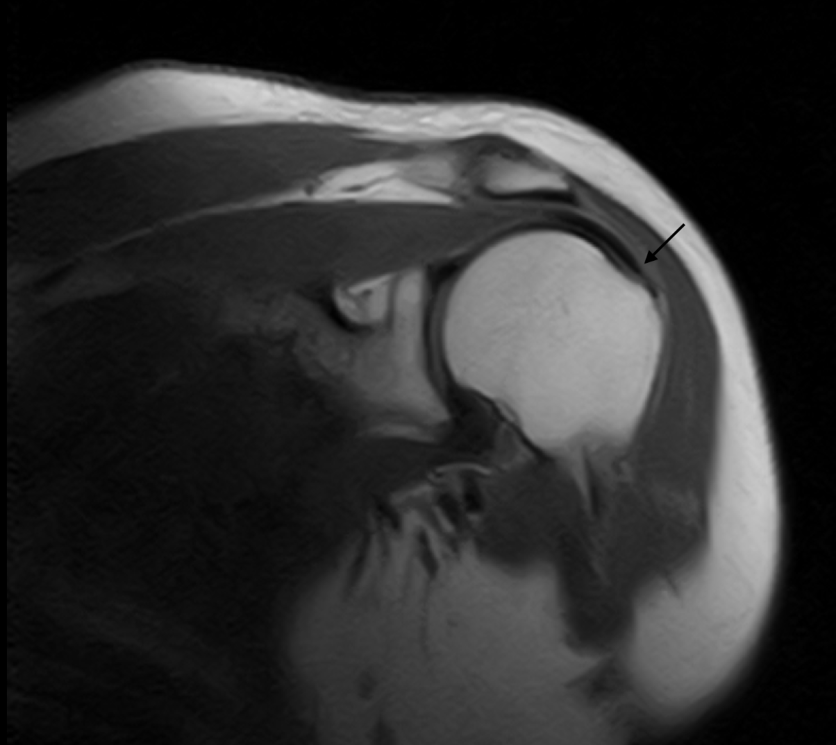
Following the clinical examination, the corresponding magnetic resonance imaging findings are reviewed below. The sequence through the supraspinatus insertion can be viewed in MRI Video 3.4 — Partial-Thickness Supraspinatus Tear.
MRI Video 3.4 — Partial-Thickness Supraspinatus TearView: Coronal oblique T1-weighted magnetic resonance imaging (MRI) through the supraspinatus tendon insertion on the greater tuberosity, demonstrating a tear involving the superficial portion of the supraspinatus tendon.
Clinical imaging obtained at Dr. Georges El Rassi Clinic, Achrafieh, Beirut, Lebanon.
MRI Figure 3.5 — Partial-Thickness Tear of the Supraspinatus Tendon
Coronal oblique T1-weighted magnetic resonance image of the supraspinatus tendon at its insertion on the greater tuberosity. The arrow indicates the tear involving the superficial portion of the supraspinatus tendon.
Clinical imaging obtained at Dr. Georges El Rassi Clinic, Achrafieh, Beirut, Lebanon.
Interpretation
In this teaching case, the patient's activity-related shoulder pain and positive provocative examination findings raised clinical concern for rotator cuff and subacromial pathology. MRI then demonstrated the corresponding partial defect involving the superficial portion of the supraspinatus tendon at the greater tuberosity. The examination and imaging are therefore interpreted together; the positive Jobe, Neer, and Hawkins-Kennedy maneuvers are supportive clinical findings rather than independent proof of the tear.
Section 2 — Total Rotator Cuff Tear
Definition
A full-thickness rotator cuff tear extends through the complete thickness of the involved tendon, creating a pathway between its articular and bursal surfaces. The defect can be focal or broad, may involve one tendon or several tendons, and can differ substantially in shape, retraction, tissue quality, and chronicity. For this reason, the words “full thickness” define the depth of the tear but do not by themselves describe its overall severity.
Tear Size and Cofield Classification
Cofield's commonly used size-based system groups full-thickness tears according to the greatest measured dimension of the tendon defect [2]. In this framework, tears are described as small when they measure less than 1 cm, medium at 1 to 3 cm, large at 3 to 5 cm, and massive when the measured defect exceeds 5 cm. Tear size is useful descriptive information, but it does not show how far the tendon has retracted or what condition the corresponding muscle is in.
Cofield Category
Greatest Tear Dimension
Small
< 1 cm
Medium
1–3 cm
Large
3–5 cm
Massive
> 5 cm
Tendon Retraction and Chronicity
Once a tendon is completely detached, its free edge may move medially away from the greater or lesser tuberosity. This tendon retraction is a separate feature from the width or length of the tear itself. With chronic tears, retraction may coexist with progressive changes in the musculotendinous unit, making assessment of tendon mobility and tissue quality increasingly relevant when planning repair [11].
Muscle Atrophy and Fatty Infiltration
Chronic cuff tears may be accompanied by loss of muscle bulk and replacement of normal muscle tissue by fat. These changes are clinically important because they reflect the condition of the muscle attached to the torn tendon and can persist even after the tendon is repaired. In longitudinal studies, enlargement of degenerative tears has been associated with progression of muscle degeneration [4], while surgical series have shown that more advanced fatty degeneration is associated with less favorable structural and functional results after repair [12].
Goutallier Classification
Goutallier and colleagues described a five-stage system for grading fatty degeneration of the rotator cuff muscles on cross-sectional imaging [3]. Grade 0 indicates no visible fat; grade 1 shows a few fatty streaks; grade 2 contains less fat than muscle; grade 3 contains approximately equal amounts of fat and muscle; and grade 4 contains more fat than muscle. This system describes muscle quality, not tear size.
Grade
Muscle Appearance
0
No visible fatty infiltration
1
Some fatty streaks within the muscle
2
Fat present, but less fat than muscle
3
Approximately equal amounts of fat and muscle
4
More fat than muscle
Massive Tears and Reparability
The term massive rotator cuff tear has not always been defined in the same way. Size-based descriptions have used a defect greater than 5 cm, whereas other shoulder literature has emphasized complete involvement of at least two cuff tendons [2][11]. “Massive,” however, does not mean the same thing as “irreparable.” Reparability is a surgical judgment that considers whether the tendon can be mobilized back to an acceptable insertion without excessive tension, together with the degree of retraction, tissue quality, chronicity, and the condition of the cuff muscles.
Clinical perspective: A complete rotator cuff tear should therefore be described in several dimensions rather than by a single label: which tendon or tendons are involved, the size and configuration of the defect, the degree of retraction, the chronicity of the lesion, and the amount of muscle atrophy or fatty infiltration. These features answer different clinical questions and should not be collapsed into one measure of “severity.”
Chapter 3 References & Primary Sources
Ellman H. Diagnosis and treatment of incomplete rotator cuff tears. Clin Orthop Relat Res. 1990;(254):64-74. doi:10.1097/00003086-199005000-00010. ࡃ Back to Text
Cofield RH. Subscapular muscle transposition for repair of chronic rotator cuff tears. Surg Gynecol Obstet. 1982;154(5):667-672. PMID:7071702. ࡃ Back to Text
Goutallier D, Postel JM, Bernageau J, Lavau L, Voisin MC. Fatty muscle degeneration in cuff ruptures. Pre- and postoperative evaluation by CT scan. Clin Orthop Relat Res. 1994;(304):78-83. doi:10.1097/00003086-199407000-00014. ࡃ Back to Text
Keener JD, Galatz LM, Teefey SA, Middleton WD, Steger-May K, Stobbs-Cucchi G, Patton R, Yamaguchi K. A prospective evaluation of survivorship of asymptomatic degenerative rotator cuff tears. J Bone Joint Surg Am. 2015;97(2):89-98. doi:10.2106/JBJS.N.00099. ࡃ Back to Text
Yamanaka K, Matsumoto T. The joint side tear of the rotator cuff. A followup study by arthrography. Clin Orthop Relat Res. 1994;(304):68-73. PMID:8020236. ࡃ Back to Text
Jobe FW, Moynes DR. Delineation of diagnostic criteria and a rehabilitation program for rotator cuff injuries. Am J Sports Med. 1982;10(6):336-339. doi:10.1177/036354658201000602. ࡃ Back to Text
Neer CS 2nd. Anterior acromioplasty for the chronic impingement syndrome in the shoulder: a preliminary report. J Bone Joint Surg Am. 1972;54(1):41-50. PMID:5054450. ࡃ Back to Text
Hawkins RJ, Kennedy JC. Impingement syndrome in athletes. Am J Sports Med. 1980;8(3):151-158. doi:10.1177/036354658000800302. ࡃ Back to Text
Teefey SA, Rubin DA, Middleton WD, Hildebolt CF, Leibold RA, Yamaguchi K. Detection and quantification of rotator cuff tears: comparison of ultrasonographic, magnetic resonance imaging, and arthroscopic findings in seventy-one consecutive cases. J Bone Joint Surg Am. 2004;86(4):708-716. doi:10.2106/00004623-200404000-00007. ࡃ Back to Text
Chun KA, Kim MS, Kim YJ. Comparisons of the various partial-thickness rotator cuff tears on MR arthrography and arthroscopic correlation. Korean J Radiol. 2010;11(5):528-535. doi:10.3348/kjr.2010.11.5.528. ࡃ Back to Text
Gerber C, Fuchs B, Hodler J. The results of repair of massive tears of the rotator cuff. J Bone Joint Surg Am. 2000;82(4):505-515. doi:10.2106/00004623-200004000-00006. ࡃ Back to Text
Goutallier D, Postel JM, Gleyze P, Leguilloux P, Van Driessche S. Influence of cuff muscle fatty degeneration on anatomic and functional outcomes after simple suture of full-thickness tears. J Shoulder Elbow Surg. 2003;12(6):550-554. doi:10.1016/S1058-2746(03)00211-8. ࡃ Back to Text
4
Biceps Pathology Snyder / Walch / Biceps Pulley
The long head of the biceps tendon (LHBT) is closely related to the superior labrum, the rotator interval, and the structures of the bicipital pulley. Because these structures function together, pathology in this region can present as a significant source of anterior shoulder symptoms and may coexist with rotator cuff disease
[1][2].
Section 1 — Biceps Tear
A Superior Labrum Anterior to Posterior (SLAP) lesion involves the superior labral region around the attachment of the long head of the biceps. Snyder and colleagues described the classic Types I through IV according to the pattern of superior labral and biceps-anchor involvement
[3].
The active compression test is one of the historically described provocative maneuvers used in the clinical assessment of superior labral and acromioclavicular pathology
[4].
Section 2 — Biceps Subluxation
Definition and Mechanical Concept
Subluxation of the long head of the biceps is a form of tendon instability in which the LHBT no longer remains normally centered within the intertubercular (bicipital) groove throughout shoulder motion. The tendon may ride onto or over the medial rim of the groove while retaining some contact with it. This differs from a true dislocation, in which the tendon loses contact with the groove and lies outside its normal osseous track
[5].
Why the Tendon Subluxates: The Bicipital Pulley
The LHBT is not stabilized by the bony groove alone. At the rotator interval and the superior entrance to the groove, the tendon is restrained by the bicipital reflection pulley, formed principally by the superior glenohumeral ligament and coracohumeral ligament in continuity with the superior border of the subscapularis
[1][6].
Disruption of this stabilizing complex can permit abnormal medial translation or subluxation of the LHBT and can be associated with subsequent tendon degeneration [6]. In Walch and colleagues’ surgical series, LHBT subluxation was closely associated with partial tearing of the subscapularis and frequently accompanied supraspinatus tearing [5].
Clinical concept: When biceps subluxation is suspected, the important question is not simply whether the biceps tendon is inflamed. The upper subscapularis, rotator interval, and bicipital pulley should also be assessed because instability often reflects failure of this stabilizing complex.
Clinical Presentation
The classic symptomatic pattern is anterior shoulder pain associated with a mechanical sensation such as painful snapping, popping, or shifting of the tendon during shoulder rotation or elevation. Athletic descriptions of LHBT subluxation emphasize tenderness over the bicipital groove and a reproducible pop during humeral rotation
[7].
In strength athletes, the same mechanical symptoms may become particularly noticeable during loaded upper-body movements that repeatedly stress the anterior shoulder. Resistance-training literature documents shoulder pain and shoulder injury as clinically important problems in weight-training athletes, while proximal biceps disorders in athletic populations are characteristically aggravated by activity
[8][9].
Examination may reproduce pain with palpation of the bicipital groove and with maneuvers that load the biceps, including resisted elbow flexion or forearm supination. Speed, Yergason, upper-cut, and related tests can support the clinical suspicion, but their diagnostic performance varies; they should therefore be interpreted together with the history and imaging rather than used as stand-alone proof of a biceps lesion
[10].
Imaging: What to Look for on Axial MRI
On axial MRI, the normal LHBT is seen as a low-signal round or oval tendon related to the bicipital groove
[11][12].
Suspicion for subluxation increases when the tendon is displaced toward or over the medial rim of the groove and when this abnormal relationship persists across sequential axial cuts; arthroscopy-correlated MRI work has specifically used displacement of the tendon over the inner rim of the bicipital groove with some remaining groove contact as an imaging criterion for subluxation
[13].
MRI assessment should also include the superior subscapularis, the superior glenohumeral ligament, the adjacent supraspinatus, and the remainder of the pulley complex
[14].
In an arthroscopy-correlated study, medial LHBT subluxation on transverse MR arthrographic images was a useful sign of a pulley lesion, while the complete diagnosis benefited from assessment of the SGHL and adjacent cuff structures [14].
Ultrasound provides an additional method of assessing the long-head biceps and can be particularly useful when instability is suspected. In a prospective comparison with arthroscopy, ultrasound demonstrated high diagnostic performance for LHBT subluxation or dislocation
[15].
Associated Tenosynovitis
Mechanical instability and tendon-sheath inflammation can coexist. MRI findings associated with bicipital tenosynovitis include abnormal tendon signal or contour and abnormal fluid or synovial change around the tendon sheath; these findings must be interpreted in the context of the complete examination because biceps-sheath fluid can accompany other shoulder pathology
[16].
The teaching case below demonstrates the relationship between the clinical history, sequential axial MRI, and an annotated image of the associated inflammatory change.
Clinical Teaching Case
Biceps Subluxation with Associated Tenosynovitis in a Bodybuilder
Patient Presentation
Clinical Presentation & History. A male recreational bodybuilder presented with pain localized predominantly to the anterior aspect of the right shoulder. The pain was provoked during heavy upper-body training, particularly movements combining shoulder elevation with elbow flexion or forearm supination. He also described an intermittent painful click or snapping sensation at the front of the shoulder during rotation under load, with improvement when training intensity was reduced. This pattern is compatible with symptomatic LHBT instability, in which anterior pain and activity-related mechanical symptoms are characteristic findings [7][9].
Training Context
Repetitive heavy resistance training involving pressing, pulling, and biceps-dominant exercises.
Pain Location
Focal anterior shoulder pain centered around the bicipital-groove region.
Mechanical Symptom
Intermittent painful snapping, clicking, or a sensation of the tendon shifting during shoulder rotation under load.
Aggravating Factors
Heavy upper-body loading, resisted elbow flexion/supination, and repeated shoulder elevation.
Clinical Correlation
The combination of anterior shoulder pain, focal bicipital-groove symptoms, and a reproducible mechanical snap raises concern for LHBT instability. Examination may demonstrate groove tenderness and may reproduce symptoms with biceps-loading maneuvers, but the diagnosis is strengthened when the clinical pattern is correlated with imaging rather than assigned from a single provocative test alone [7][10].
Interactive case media: Review the tendon across consecutive axial cuts in MRI Video 4.1, then compare it with the focused annotated still image in MRI Figure 4.2.
Axial MRI Sequence
Because biceps instability is a positional relationship between the tendon and the bicipital groove, the tendon should be followed across sequential axial images rather than judged from an isolated frame [12][13]. The following MRI sequence is presented as the imaging component of this clinical teaching case.
MRI Video 4.1 — Axial MRI of the Right Shoulder: Biceps Subluxation with Associated Tenosynovitis
Sequential axial MRI cuts of the right shoulder of a patient presenting with long-head biceps subluxation and associated tenosynovitis. The sequence allows the relationship of the LHBT to the bicipital groove and the surrounding inflammatory signal to be followed across adjacent images.
Clinical imaging obtained at Dr. Georges El Rassi Clinic, Achrafieh, Beirut, Lebanon.
The associated inflammatory component can be reviewed directly in the annotated still image below; the arrow identifies the focal bright fluid/synovial signal adjacent to the long-head biceps region. See MRI Figure 4.2 — Associated Biceps Tenosynovitis.
MRI Figure 4.2 — Arrow Demonstrating Associated Biceps Tenosynovitis
Axial MRI of the right shoulder. The arrow highlights hyperintense fluid/synovial signal adjacent to the long-head biceps region, compatible with associated tenosynovitis in the clinical context of this case.
Clinical imaging obtained at Dr. Georges El Rassi Clinic, Achrafieh, Beirut, Lebanon.
Teaching point: Biceps subluxation is the mechanical abnormality; tenosynovitis is the associated inflammatory response. They may coexist, but they should be described separately so that tendon position and tendon-sheath inflammation are not confused on imaging.
Anti-Inflammatory Procedure in This Case
The clinical procedure below is presented as part of the patient's nonoperative management. It demonstrates an ultrasound-guided corticosteroid infiltration of the subacromial–subdeltoid space through a lateral shoulder approach, used as an anti-inflammatory adjunct for associated shoulder inflammation and pain in a patient with LHBT pathology. This is a subacromial anti-inflammatory procedure rather than an injection into the substance of the biceps tendon.
Clinical Video 4.3 — Ultrasound-Guided Subacromial–Subdeltoid Corticosteroid Infiltration
Ultrasound-guided corticosteroid infiltration of the subacromial–subdeltoid space through a lateral approach over the superolateral shoulder, performed as an anti-inflammatory adjunct for associated shoulder inflammation and pain in this patient with long-head biceps pathology.
Procedure recorded at Dr. Georges El Rassi Clinic, Achrafieh, Beirut, Lebanon.
Case Interpretation
This case illustrates the value of integrating the history of anterior shoulder pain and mechanical snapping with sequential imaging of the LHBT. The MRI media demonstrate the biceps-groove relationship and the associated tendon-sheath inflammatory change, while the treatment video documents the anti-inflammatory procedure used during the patient's clinical course.
Chapter 4 References & Primary Sources
Nakata W, Katou S, Fujita A, Nakata M, Lefor AT, Sugimoto H. Biceps pulley: normal anatomy and associated lesions at MR arthrography. Radiographics. 2011;31(3):791-810. doi:10.1148/rg.313105507. ࡃ Back to Text
Virk MS, Cole BJ. Proximal biceps tendon and rotator cuff tears. Clin Sports Med. 2016;35(1):153-161. doi:10.1016/j.csm.2015.08.010. ࡃ Back to Text
Snyder SJ, Karzel RP, Del Pizzo W, Ferkel RD, Friedman MJ. SLAP lesions of the shoulder. Arthroscopy. 1990;6(4):274-279. ࡃ Back to Text
O'Brien SJ, Pagnani MJ, Fealy S, McGlynn SR, Wilson JB. The active compression test: a new and effective test for diagnosing labral tears and acromioclavicular joint abnormality. Am J Sports Med. 1998;26(5):610-613. ࡃ Back to Text
Walch G, Nové-Josserand L, Boileau P, Levigne C. Subluxations and dislocations of the tendon of the long head of the biceps. J Shoulder Elbow Surg. 1998;7(2):100-108. doi:10.1016/S1058-2746(98)90218-X. ࡃ Back to Text
Weishaupt D, Zanetti M, Tanner A, Gerber C, Hodler J. Lesions of the reflection pulley of the long biceps tendon. MR arthrographic findings. Invest Radiol. 1999;34(7):463-469. doi:10.1097/00004424-199907000-00004. ࡃ Back to Text
O'Donoghue DH. Subluxing biceps tendon in the athlete. Clin Orthop Relat Res. 1982;(164):26-29. PMID:7067297. ࡃ Back to Text
Golshani K, Cinque ME, O'Halloran P, Softness K, Keeling L, Macdonell JR. Upper extremity weightlifting injuries: diagnosis and management. J Orthop. 2018;15(1):24-27. doi:10.1016/j.jor.2017.11.005. ࡃ Back to Text
Chalmers PN, Verma NN. Proximal biceps in overhead athletes. Clin Sports Med. 2016;35(1):163-179. doi:10.1016/j.csm.2015.08.009. ࡃ Back to Text
Cardoso A, Amaro P, Barbosa L, Coelho AM, Alonso R, Pires L. Diagnostic accuracy of clinical tests directed to the long head of biceps tendon in a surgical population: a combination of old and new tests. J Shoulder Elbow Surg. 2019;28(12):2272-2278. doi:10.1016/j.jse.2019.07.007. ࡃ Back to Text
Erickson SJ, Fitzgerald SW, Quinn SF, Carrera GF, Black KP, Lawson TL. Long bicipital tendon of the shoulder: normal anatomy and pathologic findings on MR imaging. AJR Am J Roentgenol. 1992;158(5):1091-1096. doi:10.2214/ajr.158.5.1566673. ࡃ Back to Text
Nidecker A, Gückel C, von Hochstetter A. Imaging the long head of biceps tendon—a pictorial essay emphasizing magnetic resonance. Eur J Radiol. 1997;25(3):177-187. doi:10.1016/S0720-048X(97)01172-8. ࡃ Back to Text
Khil EK, Cha JG, Yi JS, Kim HJ, Min KD, Yoon YC, Jeon CH. Detour sign in the diagnosis of subluxation of the long head of the biceps tendon with arthroscopic correlation. Br J Radiol. 2017;90(1070):20160375. doi:10.1259/bjr.20160375. ࡃ Back to Text
Schaeffeler C, Waldt S, Holzapfel K, Kirchhoff C, Jungmann PM, Wolf P, Schröder M, Rummeny EJ, Imhoff AB, Woertler K. Lesions of the biceps pulley: diagnostic accuracy of MR arthrography of the shoulder and evaluation of previously described and new diagnostic signs. Radiology. 2012;264(2):504-513. doi:10.1148/radiol.12112007. ࡃ Back to Text
Armstrong A, Teefey SA, Wu T, Clark AM, Middleton WD, Yamaguchi K, Galatz LM. The efficacy of ultrasound in the diagnosis of long head of the biceps tendon pathology. J Shoulder Elbow Surg. 2006;15(1):7-11. doi:10.1016/j.jse.2005.04.008. ࡃ Back to Text
Gückel C, Nidecker A. MR arthrographic findings in tenosynovitis of the long bicipital tendon of the shoulder. Skeletal Radiol. 1998;27(1):7-12. doi:10.1007/s002560050326. ࡃ Back to Text
Disorders of the acromioclavicular (AC) joint include both acute traumatic injuries and longer-term degenerative conditions. The clinical picture therefore ranges from ligament injury after trauma to symptomatic osteoarthritis.
Section 1 — Dislocations
The Rockwood classification groups traumatic AC joint injuries into six types according to the pattern and severity of disruption [1].
Section 2 — AC Joint Inflammation
The acromioclavicular (AC) joint is a small synovial articulation that can become painful through several different mechanisms. In clinical practice, the phrase AC joint inflammation is best used as an umbrella description rather than as a single diagnosis: symptomatic inflammation may accompany primary or post-traumatic osteoarthritis, repetitive-load injury such as distal clavicle osteolysis, systemic inflammatory or crystal arthropathy, and—much less commonly—infection. Degenerative change on imaging is common and may be asymptomatic, so the diagnosis should always correlate the patient's pain pattern, focused examination, and imaging rather than relying on an isolated radiographic abnormality [4].
Major Inflammatory and Painful AC Joint Patterns
Degenerative Osteoarthritis with Reactive Synovitis
Primary AC joint osteoarthritis develops through progressive cartilage wear and subchondral change. When symptomatic, it may be accompanied by capsular irritation, joint effusion, osteophytes, and reactive bone-marrow edema. Pain is usually focal over the superior shoulder and is often aggravated by cross-body reaching, pushing, bench-press-type loading, or movements that compress the AC joint. Conservative and surgical approaches can both improve symptoms, but the available comparative evidence remains limited [5].
Post-Traumatic AC Arthritis
A previous AC sprain, separation, or direct impact can alter joint congruity and load distribution. Some patients later develop persistent focal pain and secondary degenerative inflammation even after the acute ligament injury has healed. The history of a prior injury is therefore important when evaluating chronic AC symptoms [4].
Distal Clavicle Osteolysis
Distal clavicle osteolysis is an overuse-related process classically associated with repetitive heavy loading, especially weight training. Repetitive microtrauma at the distal clavicle can produce subchondral injury, bone resorption, and AC-region pain. Patients commonly report an insidious ache over the joint that worsens with bench press, chest fly, overhead lifting, or other repetitive loading; point tenderness and pain with cross-body adduction are common examination findings [6].
Systemic Inflammatory Arthropathy
Because the AC joint is synovial, it can be involved in systemic inflammatory disease. Rheumatoid arthritis may produce AC tenderness, erosive change, tapering, osteolysis, and progressive joint destruction, particularly in patients with established systemic disease [7]. In this setting, treatment must address the systemic inflammatory disorder rather than treating the AC joint as an isolated mechanical problem.
Crystal-Induced Arthritis
Crystal arthropathy is uncommon at the AC joint but belongs in the differential diagnosis of an acutely inflamed, swollen, very tender joint. Calcium-pyrophosphate deposition disease (CPPD/pseudogout) has been documented in the AC joint, with confirmation by crystal analysis of aspirated synovial fluid [8].
Idiopathic AC Joint Inflammation
In some patients, the AC joint is clinically symptomatic and demonstrates inflammatory features despite the absence of an identifiable traumatic, repetitive-load, systemic inflammatory, crystal-related, or infectious cause. In this e-book, idiopathic AC joint inflammation is used as a descriptive diagnosis for this pattern after the major alternative causes have been considered and excluded. The diagnosis should remain clinically based, correlating focal AC-joint pain and tenderness with provocative examination findings and supportive imaging rather than relying on imaging alone. See the Clinical Teaching Case below.
Septic AC Joint Arthritis — Red Flag
Septic arthritis of the AC joint is rare but clinically important. Marked pain, warmth or swelling, systemic symptoms, immunocompromise, bacteremia risk, or unexplained acute inflammatory change should prompt urgent consideration of infection. When suspected, diagnostic aspiration/culture and appropriate antimicrobial and surgical management take priority; corticosteroid injection should not be used as routine treatment for an undiagnosed potentially infected joint [9].
Clinical Presentation
Symptomatic AC joint inflammation most often produces focal superior shoulder pain centered directly over the AC joint. The discomfort may be a persistent ache at rest and become sharper when the arm is brought across the chest, when the patient pushes through the upper extremity, performs pressing exercises, reaches to the opposite shoulder, or lies directly on the affected side. Pain that is diffuse, extends well below the elbow, or is dominated by weakness or cervical symptoms should broaden the differential diagnosis beyond the AC joint [10].
Physical Examination
Inspection: look for focal swelling, erythema, a chronic step deformity from previous AC separation, or asymmetry of the distal clavicle.
Direct palpation: reproduce the patient's familiar pain by palpating the joint line rather than the surrounding deltoid or trapezius. Local AC tenderness is sensitive but not highly specific for an injection-confirmed AC pain source [10].
Cross-Body Adduction (Scarf) Test: with the shoulder elevated to approximately 90°, the arm is passively adducted horizontally across the chest. Reproduction of the patient's focal superior AC pain is considered positive. The maneuver loads and compresses the AC joint, but it should be interpreted with the rest of the examination rather than used alone [11].
Paxinos sign: compression of the distal clavicle and acromion can reproduce AC pain and may complement other provocative maneuvers. No single special test has sufficient diagnostic strength in every clinical setting, so combinations of findings are more useful than an isolated positive maneuver [12].
Active Compression (O'Brien) test: this maneuver may be used as an adjunct when AC-joint pain is suspected, but—as with other AC provocative tests—its value is greater when interpreted together with the history, focal tenderness, and the rest of the examination rather than in isolation [12].
Complete shoulder examination: assess active and passive motion, rotator cuff strength, subacromial provocative signs, biceps/labral features, scapular mechanics, cervical screening, and neurovascular status so that concomitant or alternative sources of shoulder pain are not missed.
Clinical principle: degenerative AC findings on radiographs or MRI do not automatically prove that the AC joint is the pain generator. Concordant focal tenderness, symptom reproduction during AC-loading maneuvers, and—when uncertainty remains—a carefully performed diagnostic local-anesthetic injection can strengthen the clinical attribution [4].
Imaging
Plain radiographs remain useful for evaluating joint-space narrowing, osteophytes, distal-clavicle resorption, chronic deformity, and other osseous abnormalities. A Zanca view provides a focused radiographic assessment of the AC joint [2]. MRI is particularly useful when the diagnosis is uncertain or when associated soft-tissue and marrow abnormalities must be assessed. In symptomatic AC osteoarthritis, reported MRI features include bone-marrow edema, inferior joint distension, inferior osteophytes, and impression on the supraspinatus; bone-marrow edema was strongly associated with symptomatic disease in one comparative study [13].
Management Principles
Management is directed at the underlying cause. For noninfectious mechanical or degenerative AC pain, initial care commonly includes temporary modification of painful loading, analgesic or anti-inflammatory medication when medically appropriate, ice or other symptom-modifying measures, and rehabilitation aimed at restoring comfortable shoulder motion and scapular/shoulder mechanics [4][5]. Distal clavicle osteolysis often requires specific modification of provocative training loads [6].
A targeted AC joint injection containing local anesthetic with or without corticosteroid may be used selectively for diagnostic clarification and short-term symptom control in appropriately selected noninfectious cases. Ultrasound guidance improves the accuracy of AC joint infiltration compared with palpation-guided placement [14]. Persistent, well-localized symptomatic osteoarthritis or distal clavicle osteolysis that fails appropriate nonoperative treatment may be considered for distal clavicle excision; the classic procedure is associated with the name Mumford [3][5].
Clinical Teaching Case
Idiopathic AC Joint Inflammation
Patient Presentation
A 35-year-old male presented with pain centered over the superior aspect of the shoulder at the level of the acromioclavicular joint. The pain pattern was predominantly a localized ache around the AC region, with a sharper component during movements that compress or load the joint—particularly reaching across the body, upper-limb pushing or pressing, overhead activity, and direct pressure over the superior shoulder. This distribution is clinically compatible with an AC-joint pain generator [4].
The available case description does not establish a specific traumatic, overuse, systemic inflammatory, crystal, or infectious cause. For that reason, “idiopathic AC joint inflammation” is used here as a descriptive teaching diagnosis: it indicates symptomatic AC inflammation without an identified cause in the available clinical context, rather than a distinct formally defined disease entity.
Age
35 years.
Pain Location
Focal superior shoulder pain centered directly over the AC joint.
Pain Character
Localized aching discomfort with sharper pain during AC-loading maneuvers.
Typical Provocation
Cross-body reach, pressing/pushing, overhead loading, and direct pressure over the joint.
Physical Examination — Cross-Body Adduction Test (Scarf Test)
The Cross-Body Adduction Test (Scarf Test) is performed by elevating the patient's arm to approximately 90° and then passively moving it horizontally across the chest toward the opposite shoulder. The maneuver narrows and loads the AC articulation. The finding is clinically meaningful when it reproduces the patient's familiar focal pain directly over the AC joint, rather than nonspecific posterior shoulder tightness or diffuse pain. In isolated chronic AC lesions, the cross-body adduction stress test has shown useful sensitivity, but evidence also supports combining provocative tests and clinical findings rather than relying on this maneuver alone [11][12].
Clinical Video 5.1 — Cross-Body Adduction Test (Scarf Test)
Physical examination of the clinical teaching case. The arm is brought across the chest to load the AC joint; reproduction of the patient's localized superior AC-joint pain constitutes a positive provocative response in the appropriate clinical context.
MRI Findings
Coronal fluid-sensitive imaging is useful for evaluating the AC joint and adjacent subchondral bone. The two still images below demonstrate the AC region with high fluid-sensitive signal compatible with marrow edema/fluid and localized inflammatory activity.Such marrow and joint findings can support symptomatic AC pathology when they correspond to a concordant clinical examination [13].
A
B
MRI Figures 5.1A–B — Coronal T2-weighted / Fluid-sensitive MRI of the shoulder showing high signal intensity (bone marrow edema / fluid) and localized inflamatory changes within the acromioclavicular joint
Two coronal fluid-sensitive images from the same clinical teaching case, displayed together to emphasize the AC-joint-centered inflammatory and degenerative signal abnormalities.
MRI Video 5.2 — Full MRI Sequence of the Shoulder and AC Joint
Full MRI cut sequence provided for the clinical teaching case, allowing the AC joint and surrounding shoulder structures to be reviewed across consecutive images rather than from a single still frame.
Management in This Case
After clinical and MRI correlation localized the symptomatic process to the AC region, a targeted ultrasound-guided AC joint injection was used as the interventional component of nonoperative management. Ultrasound guidance allows real-time visualization of the joint, needle trajectory, and needle tip, and has been shown to improve the accuracy of AC joint infiltration compared with palpation-guided placement [14].
Clinical Video 5.3 — Ultrasound-guided injection targeting the acromioclavicular (AC) joint
Ultrasound-guided injection targeting the acromioclavicular (AC) joint, where the ultrasound probe is used to visualize the needle tip as it enters the joint space and delivers corticosteroid.
Teaching point: a painful AC joint is a clinical diagnosis supported—not replaced—by imaging. The most persuasive pattern is concordance between focal superior shoulder pain, reproducible AC-joint tenderness or loading pain, supportive imaging, and an etiology-appropriate response to targeted treatment. The label idiopathic should remain provisional if infection, crystal disease, systemic inflammatory arthritis, significant prior trauma, or repetitive-load osteolysis has not been adequately excluded.
Chapter 5 References & Primary Sources
Rockwood CA Jr, Williams GR, Young DC. Disorders of the acromioclavicular joint. The Shoulder. 2nd Ed. Saunders; 1998:483-553. ࡃ Back to Text
Zanca P. Shoulder pain: involvement of the acromioclavicular joint. AJR Am J Roentgenol. 1971;112(3):493-506. doi:10.2214/ajr.112.3.493. ࡃ Back to Text
Mumford EB. Resection of the marginal end of the clavicle for dislocation. J Bone Joint Surg Am. 1941;23(4):799-799. ࡃ Back to Text
Shaffer BS. Painful conditions of the acromioclavicular joint. J Am Acad Orthop Surg. 1999;7(3):176-188. doi:10.5435/00124635-199905000-00004. ࡃ Back to Text
Soler F, Mocini F, Djemeto DT, Cattaneo S, Saccomanno MF, Milano G. No differences between conservative and surgical management of acromioclavicular joint osteoarthritis: a scoping review. Knee Surg Sports Traumatol Arthrosc. 2021;29(7):2194-2201. doi:10.1007/s00167-020-06377-8. ࡃ Back to Text
Schwarzkopf R, Ishak C, Elman M, Gelber J, Strauss DN, Jazrawi LM. Distal clavicular osteolysis: a review of the literature. Bull NYU Hosp Jt Dis. 2008;66(2):94-101. PMID:18537776. ࡃ Back to Text
Petersson CJ. The acromioclavicular joint in rheumatoid arthritis. Clin Orthop Relat Res. 1987;(223):86-93. PMID:3652595. ࡃ Back to Text
Hakozaki M, Kikuchi S, Otani K, Tajino T, Konno S. Pseudogout of the acromioclavicular joint: report of two cases and review of the literature. Mod Rheumatol. 2011;21(4):440-443. doi:10.1007/s10165-011-0417-8. ࡃ Back to Text
Steinmetz RG, Maupin JJ, Smith JN, White CB. Septic arthritis of the acromioclavicular joint: a case series and review of the literature. Shoulder Elbow. 2020;12(4):272-283. doi:10.1177/1758573218815289. ࡃ Back to Text
Walton J, Mahajan S, Paxinos A, Marshall J, Bryant C, Shnier R, Quinn R, Murrell GAC. Diagnostic values of tests for acromioclavicular joint pain. J Bone Joint Surg Am. 2004;86(4):807-812. doi:10.2106/00004623-200404000-00021. ࡃ Back to Text
Chronopoulos E, Kim TK, Park HB, Ashenbrenner D, McFarland EG. Diagnostic value of physical tests for isolated chronic acromioclavicular lesions. Am J Sports Med. 2004;32(3):655-661. doi:10.1177/0363546503261723. ࡃ Back to Text
Krill MK, Rosas S, Kwon K, Dakkak A, Nwachukwu BU, McCormick F. A concise evidence-based physical examination for diagnosis of acromioclavicular joint pathology: a systematic review. Phys Sportsmed. 2018;46(1):98-104. doi:10.1080/00913847.2018.1413920. ࡃ Back to Text
Veen EJD, Donders CMD, Westerbeek RE, Derks RPH, Landman EBM, Koorevaar CT. Predictive findings on magnetic resonance imaging in patients with symptomatic acromioclavicular osteoarthritis. J Shoulder Elbow Surg. 2018;27(8):e252-e258. doi:10.1016/j.jse.2018.01.001. ࡃ Back to Text
Sabeti-Aschraf M, Lemmerhofer B, Lang S, Schmidt M, Funovics PT, Ziai P, Frenzel S, Kolb A, Graf A, Schueller-Weidekamm C. Ultrasound guidance improves the accuracy of the acromioclavicular joint infiltration: a prospective randomized study. Knee Surg Sports Traumatol Arthrosc. 2011;19(2):292-295. doi:10.1007/s00167-010-1197-y. ࡃ Back to Text
6
Calcific Tendinitis Uhthoff & Loehr
Calcific tendinitis is a cell-mediated disorder in which deposits composed largely of hydroxyapatite develop within a rotator cuff tendon [1].
Pathophysiological Phases and Interventions
Uhthoff described the condition as progressing through pre-calcific, calcific, and post-calcific stages. The calcific stage itself includes formative and resorptive phases, reflecting changes in the deposit over time [1].
Chapter 6 References & Primary Sources
Uhthoff HK, Loehr JF. Calcific tendinitis of the shoulder. Orthop Clin North Am. 1997;28(4):705-723. ࡃ Back to Text
7
Frozen Shoulder Neviaser / Zuckerman
Frozen shoulder, or adhesive capsulitis, develops through a combination of inflammation and progressive capsular tightening. Synovial inflammation is followed by fibrosis and contracture, with prominent involvement of structures such as the rotator interval and axillary recess [1].
Clinical Course and Management
Diabetes mellitus is one of the best-recognized clinical conditions associated with adhesive capsulitis [2].
Chapter 7 References & Primary Sources
Neviaser RJ, Neviaser TJ. The frozen shoulder. Clin Orthop Relat Res. 1987;(223):59-64. ࡃ Back to Text
Zuckerman JD, Rokito A. Frozen shoulder: a consensus definition. J Shoulder Elbow Surg. 2011;20(2):322-325. ࡃ Back to Text
8
Shoulder Instability Anterior / Posterior / Bipolar Bone Loss
Shoulder instability describes symptomatic loss of normal glenohumeral centering and may present as a frank dislocation, recurrent subluxation, or position-dependent apprehension. The direction of abnormal translation is clinically important because anterior and posterior instability have different injury mechanisms, characteristic soft-tissue and osseous lesions, examination findings, and reconstructive strategies.
Section 1 — Anterior Shoulder Instability
Anterior glenohumeral dislocation is the classic traumatic instability pattern of the shoulder. In the usual mechanism, the arm is forced into a vulnerable combination of abduction and external rotation, often with extension, allowing the humeral head to translate anteroinferiorly beyond the glenoid rim. The event is not simply a temporary loss of joint congruity: it can permanently deform or detach the capsulolabral stabilizers and can injure both the anterior glenoid and the posterolateral humeral head. Experimental work has shown that anterior dislocation can produce non-recoverable strain within the glenohumeral capsule, providing a biomechanical explanation for persistent laxity after the joint has been reduced [16].
Mechanism and Pathoanatomy
The principal static restraint that fails during traumatic anterior instability is commonly the anteroinferior capsulolabral complex. Detachment of the anteroinferior labrum from the glenoid rim is the classic Bankart lesion[1]. Capsular stretching or tearing may coexist with the labral lesion, and in some patients the injury extends to the glenoid rim as a bony Bankart lesion. On the humeral side, the posterolateral humeral head may impact against the anterior glenoid during dislocation, producing the characteristic Hill-Sachs lesion[2].
These lesions should be considered together rather than as isolated findings. Repeated instability can progressively enlarge anterior glenoid bone loss and humeral-head impaction damage, reducing the effective articular arc available to contain the humeral head. Clinical series of failed arthroscopic Bankart repair demonstrated that substantial glenoid deficiency and engaging Hill-Sachs lesions are powerful structural causes of recurrent instability [13]. Cadaveric work likewise showed that an anterior glenoid defect can significantly compromise anteroinferior stability even after Bankart repair [14].
Clinical Pattern and Risk of Recurrence
Patients with anterior instability often describe a traumatic event followed by pain, loss of motion, visible deformity when the shoulder is dislocated, or a subsequent sense that the shoulder may "come out" when the arm is brought into abduction and external rotation. After reduction, some shoulders become clinically stable, whereas others progress to recurrent subluxation, recurrent frank dislocation, or persistent apprehension. Age is one of the strongest clinical modifiers of recurrence: long-term prospective follow-up has shown particularly high recurrence in adolescents and young adults after a first-time traumatic anterior dislocation treated nonoperatively [12].
Typical Provocative Position
Abduction and external rotation place the anterior capsulolabral structures under tension and reproduce apprehension in many patients with traumatic anterior instability.
Soft-Tissue Failure
Bankart injury, capsular elongation or tearing, and less commonly humeral-sided capsular avulsion can reduce the effectiveness of the anterior stabilizing envelope.
Bipolar Bone Injury
Anterior glenoid bone loss and a posterolateral Hill-Sachs lesion can interact mechanically; treatment planning therefore requires assessment of both sides of the joint.
Recurrence Pattern
Recurrent events matter because repeated instability can enlarge structural defects and make a purely soft-tissue repair less reliable in a bone-deficient shoulder.
Imaging and Bone-Loss Assessment
Initial imaging confirms the direction of dislocation, excludes associated fracture, and documents the quality of the reduction. In recurrent instability, imaging must move beyond simply asking whether a Bankart or Hill-Sachs lesion is present. The clinically important questions are how much anterior glenoid articular surface has been lost, how large and how medially positioned the Hill-Sachs defect is, and whether the humeral defect can interact with the remaining glenoid during functional positions of risk. Three-dimensional CT is particularly useful for defining glenoid and humeral bone morphology, while MRI or MR arthrography can characterize the labrum, capsule, cartilage, rotator cuff, and associated soft-tissue injury.
The glenoid-track concept provides a biomechanical framework for understanding the interaction between the glenoid and a Hill-Sachs lesion. Yamamoto and colleagues mapped the zone of contact between the glenoid and the humeral head during abduction, external rotation, and horizontal extension, emphasizing that the functional significance of a humeral defect depends on its relationship to the available glenoid track rather than on humeral defect size alone [15]. This concept reinforces why anterior glenoid loss and humeral-head loss should be evaluated as a bipolar instability problem.
Lesion Characteristics and Surgical Management
Management is individualized according to whether the episode is first-time or recurrent, the patient's age and activity demands, the presence of apprehension or recurrent subluxation, the quality of the capsulolabral tissue, and the amount and interaction of glenoid and humeral bone loss. A repairable Bankart lesion in a shoulder without clinically important bone deficiency may be treated with arthroscopic capsulolabral repair when operative stabilization is indicated. In contrast, structural bone loss changes the mechanical problem: Burkhart and De Beer demonstrated markedly higher failure when significant glenoid or humeral defects were ignored during Bankart repair [13].
When anterior instability is accompanied by substantial glenoid bone loss, soft-tissue repair alone may be insufficient. The Latarjet coracoid transfer is one established reconstructive option for selected patients with clinically important anterior glenoid deficiency [3]. When a clinically important Hill-Sachs lesion contributes to instability, the humeral defect may also require treatment; the exact strategy depends on the amount of glenoid loss, the humeral defect, prior surgery, sport or occupational demand, and the overall pattern of instability. The aim is not merely to repair a visible labral tear, but to restore a stable relationship between the humeral head and the effective glenoid articular arc.
Remplissage vs Latarjet — Which Procedure Fits the Bone-Loss Pattern?
Both procedures are used to reduce recurrence in anterior shoulder instability, but they solve different mechanical problems. Remplissage primarily addresses the humeral-sided defect—the Hill-Sachs lesion—whereas the Latarjet primarily reconstructs deficient anterior glenoid bone and adds a dynamic stabilizing effect. Procedure selection therefore depends on the combined or bipolar pattern of bone loss rather than on the presence of a Hill-Sachs lesion alone [13][15].
Interactive comparison: choose a procedure below to see its principal indication, mechanism, and the clinical pattern that generally favors it.
When to Use Remplissage
Primary Indication
Recurrent anterior instability with a clinically important engaging or off-track Hill-Sachs lesion when anterior glenoid bone loss is minimal or limited. A randomized trial specifically studied patients with engaging Hill-Sachs lesions and minimal glenoid loss (<15%) and found less recurrent instability when remplissage was added to Bankart repair [19].
What It Treats
The procedure targets the posterolateral humeral-head defect rather than reconstructing the anterior glenoid rim. It is commonly performed together with arthroscopic Bankart repair when the capsulolabral lesion also requires repair.
How It Works
The posterior capsule and infraspinatus tendon are fixed into the Hill-Sachs defect, producing a capsulotenodesis that functionally converts the defect from an intra-articular engaging surface into an extra-articular lesion and reduces its ability to catch on the anterior glenoid rim during the apprehension position [18].
Think Remplissage When…
The dominant bony problem is the Hill-Sachs lesion, the anterior glenoid still provides adequate containment, and a Bankart-based soft-tissue stabilization remains mechanically reasonable.
Important limitation: remplissage is not a substitute for reconstructing a substantially deficient anterior glenoid. As glenoid bone loss increases, the risk that a soft-tissue/humeral-sided strategy will be insufficient also increases [20].
When to Use a Latarjet Procedure
Primary Indication
Anterior instability with clinically important anterior glenoid bone loss. Historically, approximately 20–25% loss has been described as the classic “critical” range for considering bony augmentation; biomechanical and clinical work has shown that failure of isolated soft-tissue stabilization may become relevant at lower levels in some patients [20].
Selected Subcritical / High-Risk Cases
Latarjet may also be selected in some patients with subcritical glenoid bone loss, particularly when the patient has high physical demands, recurrent instability, or other factors that make isolated Bankart repair less reliable. A comparative military cohort defined subcritical loss as 13.5–24% and evaluated Latarjet as an alternative to Bankart repair in this setting [22].
How It Works
The coracoid process with the attached conjoint tendon is transferred to the anteroinferior glenoid. The coracoid graft restores the deficient anterior glenoid arc, while the conjoint tendon contributes a dynamic sling effect that resists anterior translation, particularly in vulnerable arm positions [21].
Revision / High-Demand Context
The operation is also used as a revision stabilization strategy and in selected high-demand athletes with recurrent instability. Clinical series in competitive athletes have reported successful use of the Latarjet as both a primary and revision procedure [23].
Key distinction: Latarjet does not simply “fill” the Hill-Sachs lesion. Its principal reconstruction is on the glenoid side, where the transferred coracoid restores anterior bone stock and changes the mechanics of the glenohumeral articulation.
Practical Decision Guide
Hill-Sachs dominant
Engaging/off-track Hill-Sachs + minimal glenoid bone loss: Bankart repair with remplissage is a commonly used strategy because it repairs the anterior capsulolabral lesion and neutralizes the humeral defect [18][19].
Glenoid-loss dominant
Substantial anterior glenoid deficiency: a glenoid bone-restoring strategy such as Latarjet becomes more appropriate than relying on Bankart repair alone. The historically used 20–25% threshold is a useful landmark, but it should not be treated as a universal binary cutoff because biomechanical failure of soft-tissue repair has been demonstrated at lower levels [20].
Subcritical + high risk
Subcritical glenoid loss with high-demand activity, recurrent episodes, or previous stabilization failure: Latarjet may be considered depending on the complete risk profile rather than bone-loss percentage alone [22][23].
Bipolar bone loss
Meaningful defects on both sides of the joint: quantify glenoid loss and determine whether the Hill-Sachs lesion is on-track or off-track. The glenoid-track concept emphasizes that humeral and glenoid defects interact, so operative planning should address the combined geometry rather than each lesion in isolation [15].
Clinical principle: the choice is not “remplissage for every Hill-Sachs” versus “Latarjet for every recurrent dislocation.” The surgeon integrates glenoid bone loss, Hill-Sachs size and track status, recurrence pattern, tissue quality, previous surgery, age, sport, and functional demand.
Clinical Teaching Case
Anterior Shoulder Dislocation with Secondary Posterior Subluxation in the Setting of Anterior Wall Failure
Clinical Pattern
This teaching case represents a more complex pattern than an uncomplicated unidirectional anterior dislocation. The shoulder has a history of anterior instability/dislocation, but subsequent evaluation demonstrates failure of the effective anterior stabilizing wall together with posterior humeral-head subluxation. In this context, anterior wall failure refers to loss of effective anterior containment from deficient capsulolabral tissue, anterior glenoid deficiency, or failure of a previous anterior stabilization construct. Complex revision instability with loss of the anterior wall can coexist with posterior erosion, dysplasia, capsular insufficiency, or multidirectional laxity [17].
Index Direction
The initiating clinical history is anterior dislocation or recurrent anterior instability, indicating failure of the anterior stabilizing structures.
Anterior Wall Failure
The anterior capsulolabral and/or osseous containment is no longer functioning normally, so the shoulder should not be evaluated as a simple isolated Bankart lesion.
Posterior Subluxation
Posterior decentering of the humeral head indicates that instability has become bidirectional or multidirectional, or that a coexisting posterior structural abnormality is contributing to loss of centering.
Key Diagnostic Principle
Both the anterior and posterior sides of the glenohumeral joint must be examined clinically and on imaging before selecting a revision stabilization strategy.
Why Posterior Subluxation Can Appear in an Anterior-Instability Patient
Posterior subluxation is not the expected consequence of an ordinary first-time anterior dislocation. When it appears in a shoulder with previous anterior instability and anterior-wall deficiency, it should raise concern for a broader loss of glenohumeral containment. The shoulder may have capsular insufficiency in more than one direction, posterior glenoid dysplasia or erosion, altered glenoid orientation, or failure of previous reconstructive procedures. Published salvage techniques for multidirectional instability specifically describe patients with loss of the anterior wall after previous surgery together with a major posterior structural component, illustrating why apparently opposite directions of instability can coexist in the same shoulder [17].
Imaging and Mechanical Interpretation
Imaging should determine whether the anterior wall deficiency is primarily capsulolabral, osseous, or both; quantify anterior glenoid bone loss; assess the Hill-Sachs lesion; and document the degree and direction of static humeral-head subluxation. Because anterior glenoid loss reduces the effective articular arc and can compromise stability after soft-tissue repair [14], the glenoid should be assessed in three dimensions. At the same time, posterior subluxation requires evaluation of posterior labral integrity, posterior glenoid morphology, glenoid version, and capsular laxity. The glenoid-track relationship should also be considered when a Hill-Sachs lesion is present [15].
Treatment Principle
The operative plan must address the actual multidirectional structural failure rather than treating only the direction of the original dislocation. If anterior containment has failed, it may require capsulolabral reconstruction or anterior bone augmentation depending on the defect. If clinically important posterior subluxation is maintained by posterior dysplasia, erosion, labral failure, or capsular insufficiency, the posterior component must also be corrected. Complex salvage constructs combining anterior and posterior stabilization have been described for these uncommon revision patterns [17].
Teaching point: a history of anterior dislocation does not guarantee that all subsequent instability remains purely anterior. When the anterior stabilizing wall has failed and the humeral head is also subluxated posteriorly, the shoulder should be treated as a complex bidirectional or multidirectional instability problem. The cause of posterior decentering should be demonstrated clinically and radiographically rather than attributed to anterior wall failure alone.
Section 2 — Posterior Shoulder Instability
Posterior shoulder instability is less common than anterior instability and spans a spectrum from an acute traumatic posterior dislocation to recurrent posterior subluxation. Acute dislocation may follow direct trauma or a powerful internally rotating muscular contraction, including during seizure, while recurrent posterior instability may also develop through repetitive loading. In a large clinical series of acute posterior dislocations, traumatic accidents were the most frequent cause and recurrent instability developed in a clinically important minority of shoulders [4].
Provocative Position
Posterior symptoms are commonly reproduced when the shoulder is loaded in flexion, adduction, and internal rotation, a position that directs the humeral head posteriorly.
Soft-Tissue Injury
Posterior labral or capsulolabral injury may accompany traumatic dislocation and may contribute to persistent posterior translation or recurrent instability.
Osseous Injury
Posterior glenoid injury may coexist with a reverse Hill-Sachs impaction defect of the humeral head; both sides of the joint should therefore be assessed during treatment planning.
Clinical Presentation
Patients may report a definite dislocation event, posterior shoulder pain, apprehension, recurrent shifting or subluxation, mechanical symptoms, and difficulty with loaded pushing or positions that drive the humeral head posteriorly.
Reverse Hill-Sachs Lesion: How Posterior Dislocation Produces the Defect
During a posterior dislocation, the humeral head translates behind the glenoid. As the humeral head impacts against the posterior glenoid rim, an impression fracture may be created on the anteromedial aspect of the humeral head. This is the reverse Hill-Sachs lesion. It is the posterior-instability counterpart of the classic posterolateral Hill-Sachs lesion seen after anterior dislocation. MRI series of first-time traumatic posterior dislocations have demonstrated a high frequency of reverse Hill-Sachs defects and associated posterocaudal labrocapsular injury [5]. Modern three-dimensional CT analysis also localizes the reverse Hill-Sachs lesion to the anteromedial humeral head [11].
The clinical importance of the defect is not simply its presence but whether its size, depth, orientation, and relationship to the posterior glenoid permit the defect to engage the glenoid rim. Engagement can promote recurrent posterior translation and may make isolated soft-tissue stabilization insufficient. Defect assessment must therefore be integrated with posterior capsulolabral pathology, glenoid morphology or bone loss, chronicity, and the patient's functional demands [8].
Physical Examination
Examination begins with comparison of active and passive motion, scapular mechanics, generalized laxity, and direction-specific instability. Posterior translation can be assessed with posterior load-and-shift or posterior stress maneuvers, while the jerk test and Kim test are commonly used provocative tests for symptomatic posterior or posteroinferior instability [6]. The Kim test was originally described for posteroinferior labral lesions, and combining it with the jerk test improved sensitivity in the original diagnostic cohort [7]. No single examination maneuver should be interpreted in isolation; the clinically relevant finding is reproduction of the patient's pain, apprehension, posterior shift, or symptomatic clunk in a pattern consistent with the history and imaging.
Imaging
Acute posterior dislocation requires careful radiographic assessment because the injury can be missed on an isolated anteroposterior view. Axillary or equivalent orthogonal imaging is important for confirming humeral-head position. CT is useful for quantifying humeral and glenoid bone defects, while MRI evaluates the posterior capsulolabral complex, marrow edema, rotator cuff, cartilage, and associated soft-tissue injuries. In traumatic posterior dislocation, MRI may demonstrate a reverse Hill-Sachs lesion, posterior labral or capsular injury, joint effusion, and associated fracture or cuff pathology [5].
Management
Management is determined by whether the shoulder is acutely dislocated or reduced, whether instability persists, and the extent of capsulolabral and bony injury. After an acute posterior dislocation has been recognized, reduction and post-reduction stability assessment are essential. Stable shoulders with small, non-engaging reverse Hill-Sachs defects may be treated nonoperatively with protected immobilization followed by progressive rehabilitation. Recurrent symptomatic instability, a repairable posterior capsulolabral lesion that fails appropriate rehabilitation, clinically important glenoid bone loss, or a larger/engaging reverse Hill-Sachs lesion may require operative stabilization or reconstruction. Treatment algorithms are not based on one universal defect-size threshold; patient demand, chronicity, lesion size and location, engagement, and associated injuries all influence the choice of procedure [8].
Clinical / Structural Pattern
Management Principle
Reduced and stable shoulder; small non-engaging humeral defect
Protected immobilization followed by progressive rehabilitation, with clinical and imaging follow-up according to symptoms and associated injuries.
Arthroscopic posterior labral repair and capsular stabilization may be considered when symptoms persist despite appropriate nonoperative treatment.
Engaging reverse Hill-Sachs lesion
Address the humeral defect as part of stabilization. Options include reverse remplissage / modified McLaughlin-type procedures, elevation or bone graft reconstruction in selected defects, and other joint-preserving techniques based on lesion characteristics.
Very large, chronic, or non-reconstructable humeral-head defect
More extensive reconstruction or arthroplasty may be required in selected patients; the decision is individualized rather than dictated by a single imaging number.
Interactive Clinical Case
Case Report — Posterior Shoulder Fracture-Dislocation
This patient sustained a traumatic posterior shoulder dislocation / fracture-dislocation. The clinical assessment focused on whether posteriorly directed loading reproduced apprehension or a sense of instability, and imaging was used to define the associated humeral-head impaction injury before definitive treatment.
Physical Examination — Positive Posterior Apprehension Test
The examination demonstrated a positive posterior apprehension test, with posteriorly directed provocative loading reproducing the patient's apprehension in the symptomatic shoulder. This case video is provided as the physical-examination component of the teaching case. View Clinical Video 8.1 — Positive Posterior Apprehension Test. In the broader evaluation of posterior instability, this case-specific maneuver should be interpreted together with the history, posterior translation, and other validated posterior provocative maneuvers rather than as a stand-alone diagnostic test [6][7].
Clinical Video 8.1 — Positive Posterior Apprehension Test
Physical examination of the patient with traumatic posterior shoulder instability demonstrating reproduction of posterior apprehension during the provocative maneuver.
MRI Findings
MRI demonstrated an impacted humeral-head fracture with surrounding marrow edema and joint effusion. The traumatic posterior dislocation was associated with a reverse Hill-Sachs lesion, which by definition represents an impaction defect of the anteromedial humeral head created by contact with the posterior glenoid rim [5][11]. MRI also demonstrated posterior labral tearing associated with the posterior instability. Because both the posterior labral lesion and the reverse Hill-Sachs defect required stabilization, the patient underwent an arthroscopic Posterior Bankart Repair for the posterior labral tear together with reverse remplissage of the reverse Hill-Sachs lesion.
MRI Figure 8.2 — Coronal PD FS — impacted humeral head fracture with marrow edema and joint effusion
MRI from the posterior shoulder fracture-dislocation case. The associated reverse Hill-Sachs lesion represents an anteromedial humeral-head impaction defect produced during posterior dislocation.
OPERATIVE MANAGEMENT
Surgical Technique — Posterior Bankart Repair and Reverse Remplissage
Because this patient had both posterior labral tearing and a clinically significant reverse Hill-Sachs lesion, operative treatment addressed both components of the instability. The posterior labral lesion was stabilized with an arthroscopic Posterior Bankart Repair, while the humeral-head defect was treated with reverse remplissage. Arthroscopic reverse-remplissage techniques have been described to prevent an engaging reverse Hill-Sachs defect from repeatedly contacting the posterior glenoid. Published techniques include fixation of the subscapularis tendon and middle glenohumeral ligament (MGHL) into the reverse Hill-Sachs defect to convert the humeral defect into an extra-articular lesion [9][10]. The following steps document the operative management used in this patient.
1 · Posterior Bankart Repair
The torn posterior labrum was repaired arthroscopically to restore the posterior capsulolabral restraint and stabilize the posterior glenoid-labral complex.
2 · Prepare the Humeral Defect
The reverse Hill-Sachs defect was prepared arthroscopically using a shaver to create a clean recipient surface for soft-tissue fixation.
3 · Reverse Remplissage into the Defect
A reverse remplissage was performed using the subscapularis tendon and the middle glenohumeral ligament. In this case, a tenodesis suture was passed through these structures and secured into the reverse Hill-Sachs defect.
4 · Closure & Immobilization
Arthroscopic portals were closed with nylon sutures and sterile dressings were applied. The shoulder was immobilized in abduction with neutral rotation. The patient tolerated the procedure and was transferred to recovery in stable condition.
Rationale
Reverse remplissage fills the intra-articular impaction defect with soft tissue so that the defect behaves functionally as an extra-articular lesion and is less able to engage the posterior glenoid rim, avoiding an open bone-grafting procedure in appropriately selected defects [9][10].
Operative Figure 8.3 — Reverse Hill-Sachs Defect with the Subscapularis Tendon and Middle Glenohumeral Ligament Positioned for Remplissage
Reverse Hill-Sachs defect with the subscapularis tendon and middle glenohumeral ligament positioned for remplissage.
Taken during the procedure at Saint George Hospital, Achrafieh.
Operative Figure 8.4 — Tenodesis Suture Secured into the Reverse Hill-Sachs Defect
Tenodesis suture secured into the reverse Hill-Sachs defect, completing the remplissage.
Taken during the procedure at Saint George Hospital, Achrafieh.
Teaching point: posterior dislocation creates a characteristic injury pattern that differs from anterior instability. The reverse Hill-Sachs lesion is located on the anteromedial humeral head; when the defect is clinically significant or engaging, successful stabilization may require treatment of both the posterior capsulolabral pathology and the humeral-head defect.
Chapter 8 References & Primary Sources
Bankart ASB. The pathology and treatment of recurrent dislocation of the shoulder-joint. Br J Surg. 1938;26(101):23-29. doi:10.1002/bjs.18002610104. ࡃ Back to Text
Hill HA, Sachs MD. The grooved defect of the humeral head: a frequently unrecognized complication of dislocations of the shoulder joint. Radiology. 1940;35(6):690-700. doi:10.1148/35.6.690. ࡃ Back to Text
Latarjet M. Treatment of recurrent dislocation of the shoulder. Lyon Chir. 1954;49(8):994-997. PMID:13234709. ࡃ Back to Text
Robinson CM, Seah M, Akhtar MA. The epidemiology, risk of recurrence, and functional outcome after an acute traumatic posterior dislocation of the shoulder. J Bone Joint Surg Am. 2011;93(17):1605-1613. doi:10.2106/JBJS.J.00973. ࡃ Back to Text
Saupe N, White LM, Bleakney R, Schweitzer ME, Recht MP, Jost B, et al. Acute traumatic posterior shoulder dislocation: MR findings. Radiology. 2008;248(1):185-193. doi:10.1148/radiol.2481071003. ࡃ Back to Text
Tannenbaum E, Sekiya JK. Evaluation and management of posterior shoulder instability. Sports Health. 2011;3(3):253-263. doi:10.1177/1941738111400562. ࡃ Back to Text
Kim SH, Park JS, Jeong WK, Shin SK. The Kim test: a novel test for posteroinferior labral lesion of the shoulder—a comparison to the jerk test. Am J Sports Med. 2005;33(8):1188-1192. doi:10.1177/0363546504272687. ࡃ Back to Text
van der List JP, Waterman BR. Humeral head reconstruction of reverse Hill-Sachs lesions. Instr Course Lect. 2024;73:559-571. PMID:38090925. ࡃ Back to Text
Duey RE, Burkhart SS. Arthroscopic treatment of a reverse Hill-Sachs lesion. Arthrosc Tech. 2013;2(2):e155-e159. doi:10.1016/j.eats.2013.01.007. ࡃ Back to Text
Lavender CD, Hanzlik SR, Pearson SE, Caldwell PE 3rd. Arthroscopic reverse remplissage for posterior instability. Arthrosc Tech. 2016;5(1):e43-e47. doi:10.1016/j.eats.2015.09.005. ࡃ Back to Text
Yang K, Yamamoto N, Takahashi N, Kamijo H, Okamura K, Mihata T, et al. Location and size of the reverse Hill-Sachs lesion in patients with traumatic posterior shoulder instability. J Shoulder Elbow Surg. 2025;34(1):88-95. doi:10.1016/j.jse.2024.03.017. ࡃ Back to Text
Hovelius L, Olofsson A, Sandström B, Augustini BG, Krantz L, Fredin H, et al. Nonoperative treatment of primary anterior shoulder dislocation in patients forty years of age and younger: a prospective twenty-five-year follow-up. J Bone Joint Surg Am. 2008;90(5):945-952. doi:10.2106/JBJS.G.00070. ࡃ Back to Text
Burkhart SS, De Beer JF. Traumatic glenohumeral bone defects and their relationship to failure of arthroscopic Bankart repairs: significance of the inverted-pear glenoid and the humeral engaging Hill-Sachs lesion. Arthroscopy. 2000;16(7):677-694. doi:10.1053/jars.2000.17715. ࡃ Back to Text
Itoi E, Lee SB, Berglund LJ, Berge LL, An KN. The effect of a glenoid defect on anteroinferior stability of the shoulder after Bankart repair: a cadaveric study. J Bone Joint Surg Am. 2000;82(1):35-46. doi:10.2106/00004623-200001000-00005. ࡃ Back to Text
Yamamoto N, Itoi E, Abe H, Minagawa H, Seki N, Shimada Y, et al. Contact between the glenoid and the humeral head in abduction, external rotation, and horizontal extension: a new concept of glenoid track. J Shoulder Elbow Surg. 2007;16(5):649-656. doi:10.1016/j.jse.2006.12.012. ࡃ Back to Text
Rainis CA, Browe DP, McMahon PJ, Debski RE. Capsule function following anterior dislocation: implications for diagnosis of shoulder instability. J Orthop Res. 2013;31(6):962-968. doi:10.1002/jor.22300. ࡃ Back to Text
de Cabo G, Ramos-Murillo P, González-Martín D, Álvarez-Benito N, Poyato-Núñez F, Campesino-Nieto SV, et al. Anterior arthroscopic approach for multidirectional shoulder instability: posterior bone block, dynamic anterior stabilization, and modified McLaughlin. Arthrosc Tech. 2024;13(11):103115. doi:10.1016/j.eats.2024.103115. ࡃ Back to Text
Purchase RJ, Wolf EM, Hobgood ER, Pollock ME, Smalley CC. Hill-Sachs “remplissage”: an arthroscopic solution for the engaging Hill-Sachs lesion. Arthroscopy. 2008;24(6):723-726. doi:10.1016/j.arthro.2008.03.015. ࡃ Back to Text
MacDonald P, McRae S, Old J, Marsh J, Dubberley J, Stranges G, et al. Arthroscopic Bankart repair with and without arthroscopic infraspinatus remplissage in anterior shoulder instability with a Hill-Sachs defect: a randomized controlled trial. J Shoulder Elbow Surg. 2021;30(6):1288-1298. doi:10.1016/j.jse.2020.11.013. ࡃ Back to Text
Shin SJ, Koh YW, Bui C, Jeong WK, Akeda M, Cho NS, et al. What is the critical value of glenoid bone loss at which soft tissue Bankart repair does not restore glenohumeral translation, restricts range of motion, and leads to abnormal humeral head position? Am J Sports Med. 2016;44(11):2784-2791. doi:10.1177/0363546516656367. ࡃ Back to Text
Yamamoto N, Muraki T, An KN, Sperling JW, Cofield RH, Itoi E, et al. The stabilizing mechanism of the Latarjet procedure: a cadaveric study. J Bone Joint Surg Am. 2013;95(15):1390-1397. doi:10.2106/JBJS.L.00777. ࡃ Back to Text
Min KS, Wake J, Cruz C, Miles R, Chan S, Shaha J, et al. Surgical treatment of shoulder instability in active-duty service members with subcritical glenoid bone loss: Bankart vs. Latarjet. J Shoulder Elbow Surg. 2023;32(4):771-775. doi:10.1016/j.jse.2022.10.011. ࡃ Back to Text
Rossi LA, Bertona A, Tanoira I, Maignon GD, Bongiovanni SL, Ranalletta M. Comparison between modified Latarjet performed as a primary or revision procedure in competitive athletes: a comparative study of 100 patients with a minimum 2-year follow-up. Orthop J Sports Med. 2018;6(12):2325967118817233. doi:10.1177/2325967118817233. ࡃ Back to Text