The Shoulder
From Clinic to Recovery
An advanced interactive reference in shoulder pathologies, arthroscopy, and rehabilitation — anatomy through operative technique to return-to-play, cited to source.
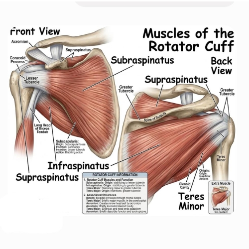
The glenohumeral joint balances extreme mobility with intrinsic instability1. The shallow glenoid fossa accommodates less than one-third of the humeral head surface area, making dynamic stabilization through the rotator cuff critical for function1.
Axillary N. (C5-C6): teres minor & deltoid1.
Structural foundations
Rotator cuff musculature specifications
| Muscle | Scapular origin | Humeral insertion | Primary action | Innervation |
|---|---|---|---|---|
| Supraspinatus | Supraspinous fossa | Superior facet, greater tubercle | Initiates abduction; centres head | Suprascapular N. (C5-C6) |
| Infraspinatus | Infraspinous fossa | Middle facet, greater tubercle | External rotation; posterior stability | Suprascapular N. (C5-C6) |
| Teres minor | Lateral scapular border | Inferior facet, greater tubercle | External rotation in elevation | Axillary N. (C5-C6) |
| Subscapularis | Subscapular fossa | Lesser tubercle | Internal rotation; anterior restraint | Upper/lower subscapular N. (C5-C6) |
Coracohumeral & glenohumeral ligamentous complex
The coracohumeral ligament (CHL) originates from the base of the coracoid process and spans across the rotator interval, enveloping the supraspinatus tendon margin1. It acts in conjunction with the superior glenohumeral ligament (SGHL) to form a stabilizing pulley system for the long head of the biceps tendon within the bicipital groove3.
Concavity-compression mechanism
Because the osseous socket is inherently flat, stability is maintained by pressing the convex humeral head into the concave glenoid socket bounded by the glenoid labrum1. Dynamic co-contraction of the rotator cuff creates compressive forces preventing translation under load1.
Mechanical compression of subacromial structures against the coracoacromial arch during overhead arm elevation2. Morphological variations of the acromion directly constrain the outlet space for the supraspinatus tendon6.
Clinical pathway
Pathophysiology & classification
Subacromial impingement involves dynamic or mechanical compression of the subacromial bursa and supraspinatus tendon beneath the coracoacromial arch2. Bigliani classified acromial morphology into three types: Type I (flat, 17%), Type II (curved, 43%), and Type III (hooked, 39%), with Type III showing the highest association with full-thickness rotator cuff tears6.
Physical examination & diagnostics
- Neer sign: positive pain response on passive forced forward flexion with the arm internally rotated2.
- Hawkins-Kennedy test: pain elicited by passive internal rotation at 90° forward flexion.
- Jobe (empty can) test: pain or weakness during resisted elevation in the scapular plane with thumbs pointed down.
In-office subacromial infiltration
Technique: posterolateral approach. Needle inserted 1-2 cm inferior to the posterolateral border of the acromion, directed toward the undersurface of the acromion tip.
Injectate mix: 5 mL 1% lidocaine + 40 mg methylprednisolone or triamcinolone acetonide.
Arthroscopic subacromial decompression (SAD)
Under arthroscopic visualization from the posterior portal, a lateral working portal is established2. A high-speed burr resects the anteroinferior acromial spur, converting a Type II/III acromion into a flat Type I arch alongside subacromial bursectomy2.
Rehabilitation protocol
- Phase I (weeks 0-2): sling for comfort, early PROM and active-assisted ROM.
- Phase II (weeks 2-6): full AROM, scapular stabilization, rotator cuff isometric strengthening.
- Phase III (weeks 6+): progressive resistance training and sports-specific functional conditioning.
A spectrum ranging from tendinopathy and partial-thickness tears to massive full-thickness retractions with fatty degeneration7.
Clinical pathway
Pathophysiology & classification
Rotator cuff tears stem from intrinsic age-related hypovascular degeneration and extrinsic mechanical impingement1. Progression leads to muscle atrophy and irreversible fatty infiltration, graded 0-4 using the Goutallier CT/MRI classification7.
Physical examination & imaging
- Drop-arm sign: inability to smoothly decelerate the arm from 90° passive abduction.
- External rotation lag sign: inability to hold the shoulder in full external rotation (infraspinatus).
- Belly-press test: assesses subscapularis power.
In-office infiltration
Ultrasound-guided diagnostic subacromial injection: 4 mL 1% lidocaine + 40 mg Depo-Medrol to differentiate pain-inhibited strength from true structural disruption.
Arthroscopic double-row suture bridge repair
The footprint at the greater tubercle is debrided down to bleeding bone1. Medial suture anchors are placed at the articular margin; suture tails are passed through the tendon footprint and secured laterally with sutureless anchors to restore anatomic footprint contact area1.
Postoperative rehabilitation
- Weeks 0-6: abduction sling immobilization. PROM limited to 120° elevation and 30° external rotation.
- Weeks 6-12: discontinue sling; initiate AAROM and active ROM.
- Weeks 12+: progressive resistive strengthening; avoid full loading until 6 months.
Pathology of the superior labrum anterior to posterior (SLAP) and instability or tendinopathy of the long head of biceps tendon (LHBT)4.
Clinical pathway
Anatomy & Snyder classification
Snyder classified superior labral tears into Types I-IV4. Type II (fraying with detachment of the biceps anchor from the superior glenoid tubercle) is the most common clinical variant4.
Physical exam & diagnostics
- O'Brien test: deep joint pain with resisted elevation in 10° adduction/internal rotation, relieved by supination8.
- Speed's test: pain in the bicipital groove with resisted elevation and the forearm supinated.
- Yergason's test: pain during resisted supination/external rotation with the elbow flexed 90°3.
In-office bicipital groove infiltration
High-frequency linear ultrasound guidance targeting the bicipital sheath inside the bicipital groove: 3 mL 1% lidocaine + 20 mg Depo-Medrol.
Arthroscopic biceps tenodesis / tenotomy
The tendon is released from the superior glenoid anchor and fixed extra-articularly into the bicipital groove or subpectoral region using an interference screw or suture anchor to avoid post-op stiffness3.
Rehabilitation guidelines
- Weeks 0-4: sling support; avoid active elbow flexion and forearm supination.
- Weeks 4-8: active elbow flexion without load; gradual restoration of full ROM.
- Weeks 8+: progressive biceps dynamic resistance strengthening.
Degenerative arthrosis, post-traumatic osteolysis, and acute dislocation of the acromioclavicular (AC) and coracoclavicular (CC) ligament complex1.
Clinical pathway
Pathophysiology & classification
Rockwood classification: Type I (sprain), Type II (AC disruption, CC intact), Type III (100% distal clavicle displacement), Type IV (posterior displacement into trapezius), Type V (>100% displacement), Type VI (inferior displacement beneath the coracoid)1.
Physical examination & imaging
- Step-off deformity: palpable elevation of the distal clavicle.
- Cross-body adduction test: pain over the superior AC joint line during horizontal adduction.
- Paxinos test: thumb pressure on the posterior acromion + index pressure on the distal clavicle reproduces pain.
In-office AC joint infiltration
Palpate the superior notch between clavicle and acromion; 25-gauge needle inserted superior-to-inferior: 1.5 mL 1% lidocaine + 20 mg triamcinolone acetonide.
Surgical reconstruction
Indicated for high-grade Rockwood IV/V/refractory III. Reconstruction uses cortical fixation button devices (e.g., TightRope) or a tendon graft passed through clavicle and coracoid bone tunnels, alongside distal clavicle resection (Mumford procedure, 5-8 mm) if degenerate1.
Rehabilitation protocol
- Weeks 0-6: sling support to reduce vertical clavicular shear forces.
- Weeks 6-12: gentle ROM restoration below 90° elevation; progressive trapezius/deltoid activation.
- Weeks 12+: return to full overhead function and sport activities.
Cell-mediated calcium hydroxyapatite deposition within the rotator cuff tendons, driven by local tissue hypoxia and metaplasia5.
Clinical pathway
Uhthoff pathophysiological phases
Physical exam & diagnostics
- Guarding: severe limitation of active and passive movements due to acute bursal pressure.
- Tenderness: exquisite tenderness on direct palpation over the anterior greater tubercle.
US-guided needle barbotage & infiltration
Under real-time ultrasound guidance, two 18-gauge needles penetrate the calcific deposit. Repeated saline lavage aspirates the fluid calcium deposit. The subacromial bursa is then injected with 40 mg methylprednisolone to manage secondary bursitis.
Arthroscopic evacuation
Indicated for refractory chronic pain. The tendon is identified, probed, and incised parallel to its fibers to evacuate calcific material5. The cavity is irrigated thoroughly; residual defects are repaired with suture anchors if necessary.
Rehabilitation protocol
- Days 1-7: early passive-assisted pendulum exercises as tolerated.
- Weeks 2-4: full active ROM and progressive resistance strengthening as the inflammatory phase settles.
Fibroblastic proliferation and capsular contracture, localized predominantly in the rotator interval and axillary recess9.
Clinical pathway
Pathophysiology & stages
Cytokine-mediated inflammatory synovitis leading to capsular fibrosis (elevated TGF-beta)9. Phases: 1. Freezing (painful), 2. Frozen (stiff), 3. Thawing9.
Physical examination & imaging
- Global loss of ROM: equal restriction of both active and passive range of motion, most notably external rotation with the arm at the side (< 15°)1.
- Scapular hike: early compensatory scapulothoracic elevation during abduction attempts.
Glenohumeral hydrodilatation & infiltration
Ultrasound-guided posterior glenohumeral injection: 40 mg triamcinolone acetonide + 4 mL 1% lidocaine + 15-20 mL normal saline to distend the capsule and rupture adhesions.
Manipulation under anesthesia (MUA) & release
If conservative therapy fails after 6 months. Controlled, sequential manipulation into flexion, abduction, and rotation, combined with arthroscopic 360° capsular release (releasing the rotator interval, coracohumeral ligament, and inferior capsule)9.
Rehabilitation guidelines
- Day 1 post-op: immediate intensive physical therapy within 24 hours to preserve capsular volume.
- Weeks 1-6: daily stretching routines targeting end-range external rotation and elevation.
Traumatic anterior glenohumeral dislocation resulting in labral detachment and bone loss10.
Clinical pathway
Pathophysiology & lesions
Physical examination & diagnostics
- Apprehension test: anxiety and resistance when the shoulder is brought to 90° abduction and full external rotation.
- Jobe relocation test: reduction of apprehension when a posterior force is applied to the anterior proximal humerus.
In-office infiltration
Diagnostic intra-articular lidocaine injection (5 mL 1%) used to differentiate true mechanical instability from painful rotator cuff weakness prior to rehabilitation.
Arthroscopic Bankart vs. open Latarjet
If glenoid bone loss is < 15%, an arthroscopic Bankart repair is performed using suture anchors placed at the anteroinferior rim (3 to 6 o'clock)10. If bone loss is > 15-20%, an open Latarjet procedure is performed (coracoid process transfer with the short head of biceps to the anterior glenoid rim)10.
Rehabilitation protocol
- Weeks 0-4: sling immobilization. Restrict external rotation beyond neutral (0°) to protect the anterior repair.
- Weeks 4-8: gradual increase in external rotation (up to 30°) and elevation.
- Months 3-6: plyometrics and sports-specific functional training; return to contact play at 6 months.
Primary Academic Citations
Vancouver-style literature index · click any superscript in the text to jump here.