The Scorpion/Spider Remplissage Technique for Anterior Instability With Multidirectional Capsular Laxity
A three-stitch, humeral-sided capsulotenodesis designed to address a Hill-Sachs defect while incorporating a posteroinferior capsular shift in high-demand patients with multidirectional instability.
Abstract
Background: Recurrent instability after arthroscopic shoulder stabilization may result from the combined effects of bipolar bone loss and capsular laxity. The Scorpion/Spider technique was developed to combine Hill-Sachs defect filling with humeral-sided capsular tightening.
Technique: The technique uses a three-stitch remplissage construct consisting of a deep central stitch, a superior oblique stitch, and a posteroinferior capsular-shift stitch. A 5.5-mm triple-loaded anchor is placed in the deepest portion of the Hill-Sachs defect. The sutures are individually identified and tied under direct subacromial visualization after standard anteroinferior capsulolabral repair.
Conclusion: The Scorpion/Spider technique may be considered for selected patients with anterior instability, a substantial Hill-Sachs defect, and multidirectional capsular laxity. Its clinical effectiveness and safety require prospective validation.
Introduction
Recurrence following arthroscopic shoulder stabilization is influenced by patient age, activity level, capsulolabral injury, glenoid bone loss, humeral bone loss, capsular laxity, and anatomical variants in the shape, size, direction, and depth of both the native glenoid and the humeral head. Hill-Sachs lesions should therefore be assessed in relation to the glenoid rather than according to size alone [1]. A large Hill-Sachs lesion has itself been shown to be significantly associated with postoperative recurrence, underscoring that humeral-sided bone loss is a relevant risk factor independent of the glenoid [2].
The remplissage procedure fills the Hill-Sachs defect with the posterior capsule and infraspinatus tendon. It is commonly performed with Bankart repair in selected patients with engaging or large Hill-Sachs lesions and has been associated with improved stability outcomes [3].
Multidirectional instability represents a different biomechanical problem because it involves symptomatic instability in more than one direction and is commonly associated with capsular redundancy. Treatment may require reduction of excessive capsular volume in addition to repair of the anterior labrum [4]. Generalized shoulder hyperlaxity has been identified as a significant risk factor for recurrent instability after Bankart repair, reinforcing the rationale for directly addressing capsular redundancy rather than the anterior labrum alone [5]. Historically, an inferior capsular shift has been described as an effective open technique for addressing this redundancy, and its underlying principle of volume reduction informs arthroscopic capsular-shift constructs today [6].
The Scorpion/Spider technique was developed to address these combined findings by adding a posteroinferior humeral-sided capsular shift to a multi-stitch remplissage construct.
Indications and Preoperative Planning
Potential indications include recurrent anterior instability associated with a Hill-Sachs defect and arthroscopic evidence of anterior and posterior capsular laxity, particularly after failure of previous soft-tissue stabilization.
Preoperative assessment should include a history of instability episodes, physical examination in multiple directions, evaluation of generalized laxity, and three-dimensional imaging of the glenoid and humeral head. A structured evaluation for multidirectional instability, including assessment of voluntary versus involuntary components and generalized ligamentous laxity, further refines patient selection [7].
The glenoid-track method evaluates the relationship between the Hill-Sachs interval and the available glenoid track. When the Hill-Sachs interval exceeds the glenoid track, the lesion is classified as off-track and has an increased risk of engagement [8].
This technique should not be used to compensate for critical glenoid bone loss when a bone-augmentation procedure is indicated.
Inclusion criteria: Patients with recurrent anterior shoulder instability, clinical evidence of multidirectional capsular laxity (e.g., a positive drive-through sign, generalized hyperlaxity), and a large engaging or off-track Hill-Sachs lesion following failed conservative management were considered candidates for this technique.
Imaging thresholds and lesion measurements: Three-dimensional CT reconstruction was used to calculate the Hill-Sachs interval (HSI) and glenoid track width. Surgery was indicated when the HSI exceeded the available glenoid track, per the glenoid-track method described above.
Surgical Technique
The complete arthroscopic procedure is demonstrated in the accompanying video (Video 1).
-
Position and diagnostic assessment.
Place the patient in the beach-chair position with slight traction. During diagnostic arthroscopy, assess anterior and posterior translation. A positive drive-through sign may indicate capsular laxity. If the arthroscope passes easily across the joint during this assessment, this is a further indicator of underlying shoulder laxity.
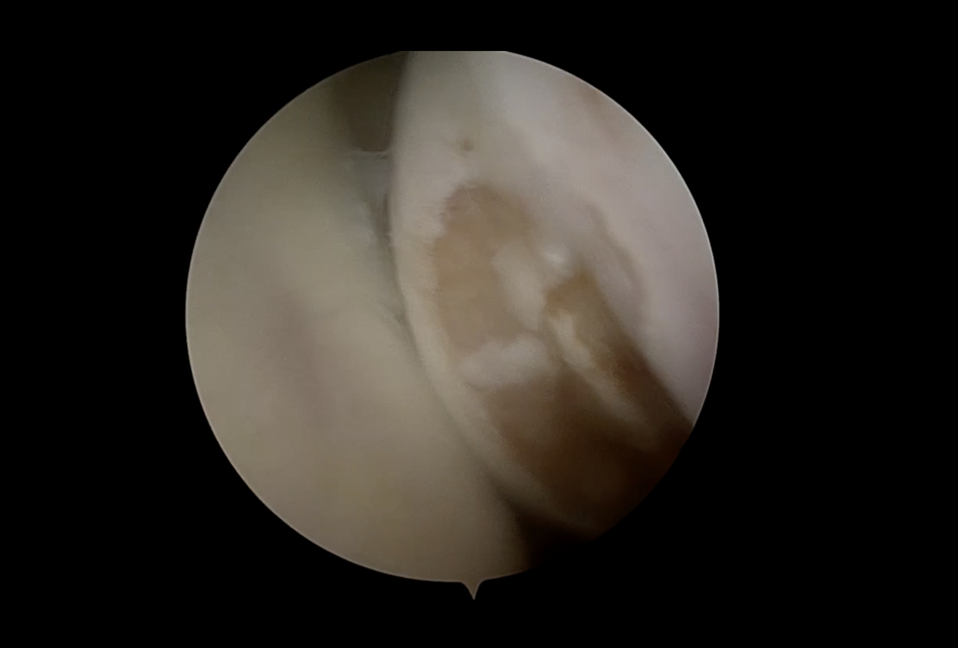
Figure 1. Arthroscopic view of the Hill-Sachs lesion on the posterior humeral head observed during diagnostic assessment. -
Accessory portal.
Establish an anterior accessory portal according to the location of the Hill-Sachs lesion. Use a spinal needle to confirm the appropriate trajectory.
-
Defect preparation.
Place the arm in internal rotation. The assistant applies an anterior drawer force to the arm to maintain joint space throughout preparation. Use a shaver and burr without a cannula to remove scar tissue and prepare a stable bony bed. Avoid excessive debridement.
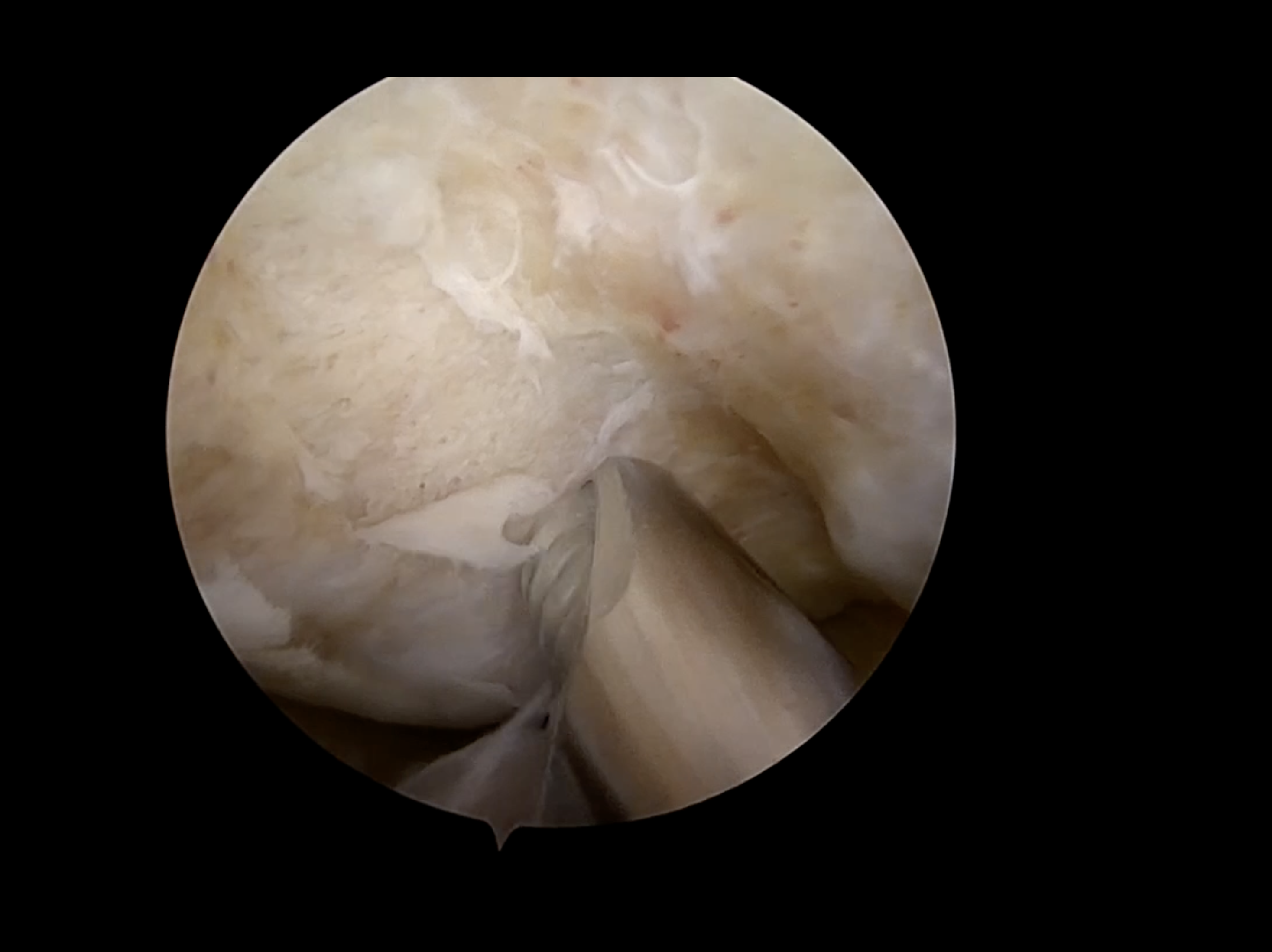
Figure 2. Bony preparation of the Hill-Sachs lesion for the subsequent remplissage. -
Anchor placement.
Depending on the size of the defect and the degree of laxity, use one or two 5.5-mm triple-loaded anchors. Prepare the bone with a screwdriver (size 4.5) and place the anchor in the deepest portion of the Hill-Sachs lesion. Confirm cartilage clearance across two arthroscopic views before proceeding.
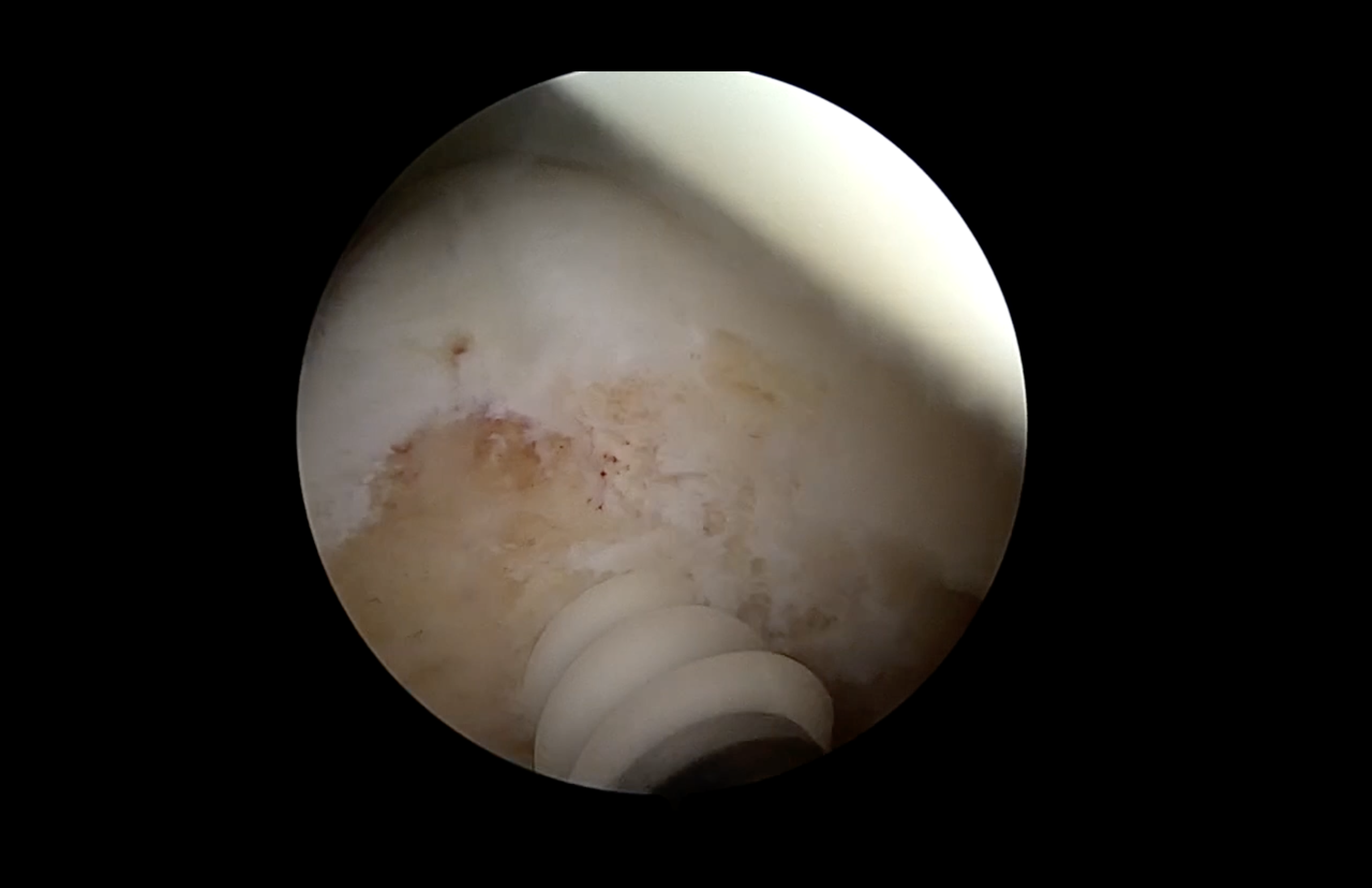
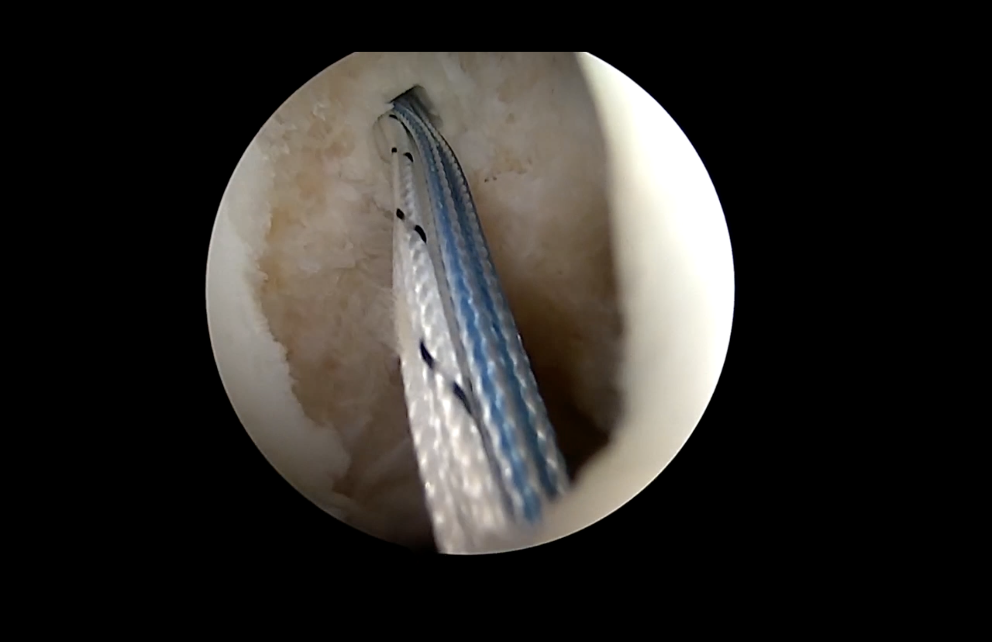
Figure 3 (A–B). Stepwise insertion of a triple-loaded 5.5-mm anchor into the deepest portion of the Hill-Sachs lesion, with confirmation of secure seating and cartilage clearance. -
Deep central stitch (the black and white suture).
Using a BirdBeak suture passer (straight or anteriorly curved), place one bite adjacent to the humeral head in the same plane as the anchor and a second bite approximately 1–1.5 cm from the anchor entry hole. This stitch is intended to compress the capsule and infraspinatus into the defect.
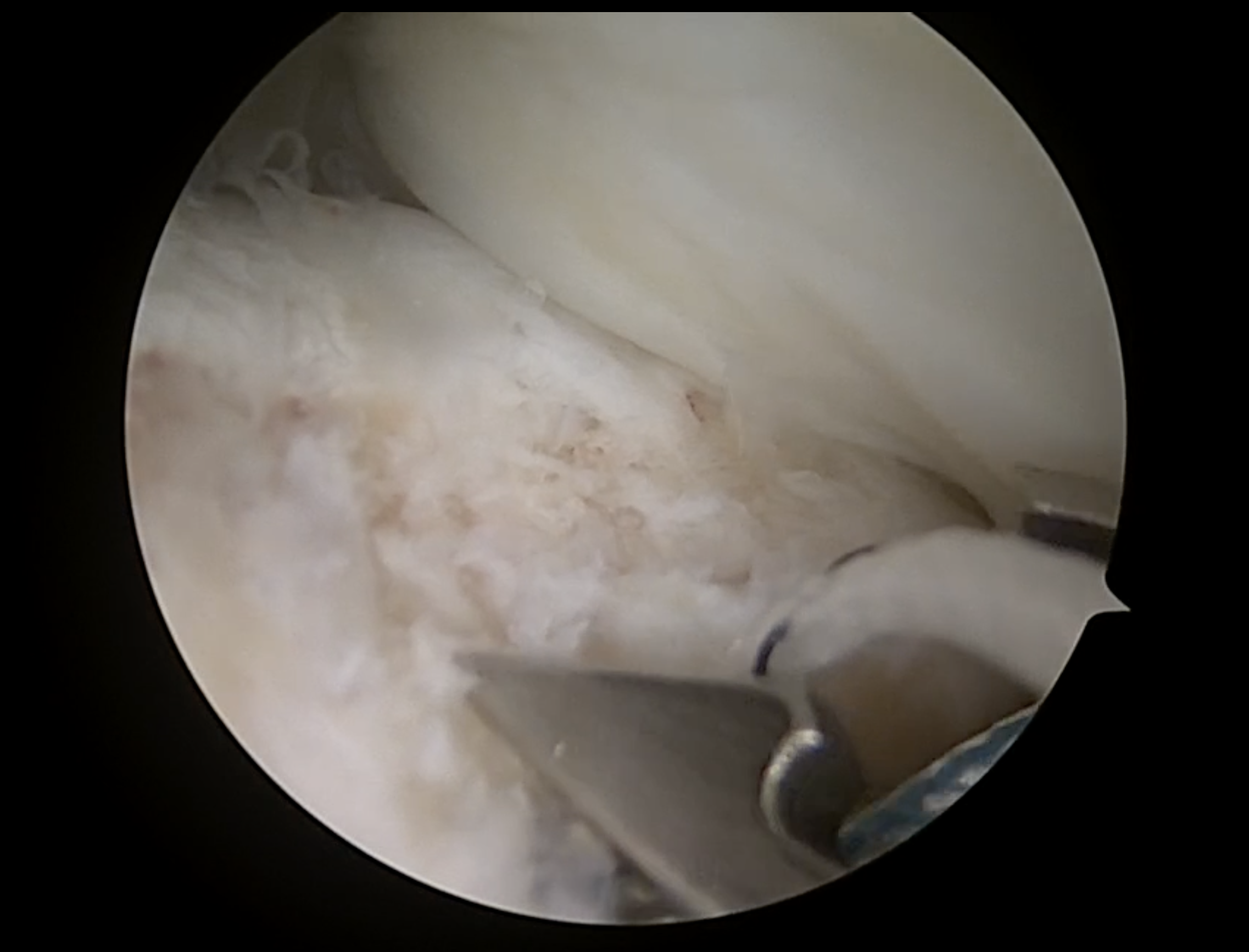
Figure 4. Passing of the deep central stitch adjacent to the humeral head. -
Superior oblique stitch (the white suture).
Pass a second stitch through the infraspinatus in a superior-to-inferior oblique direction to cover the superior and anterior portions of the defect.
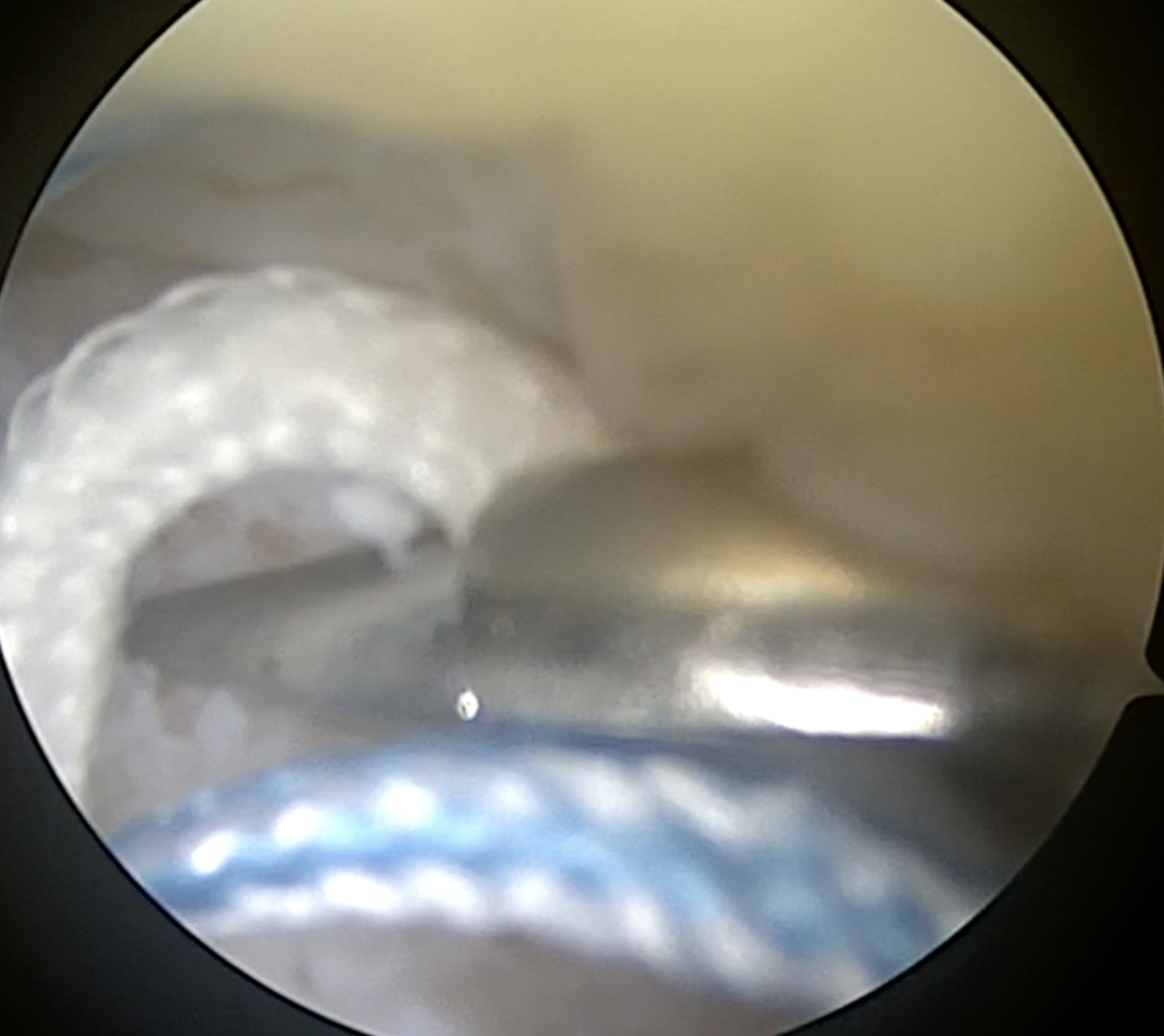
Figure 5. Shuttling of the superior oblique stitch. -
Posteroinferior capsular-shift stitch (blue one).
With the arm in maximal internal rotation, pass the third stitch through the inferior capsule approximately 1–2 cm inferior to the anchor, depending on the degree of posteroinferior laxity. This stitch reproduces, on the humeral side, the posteroinferior capsular-shift principle described in the Jobe technique.
-
Bankart repair.
After passage of the remplissage sutures, perform the standard anteroinferior capsulolabral reconstruction, with anchor placement on the anterior aspect of the glenoid labrum.
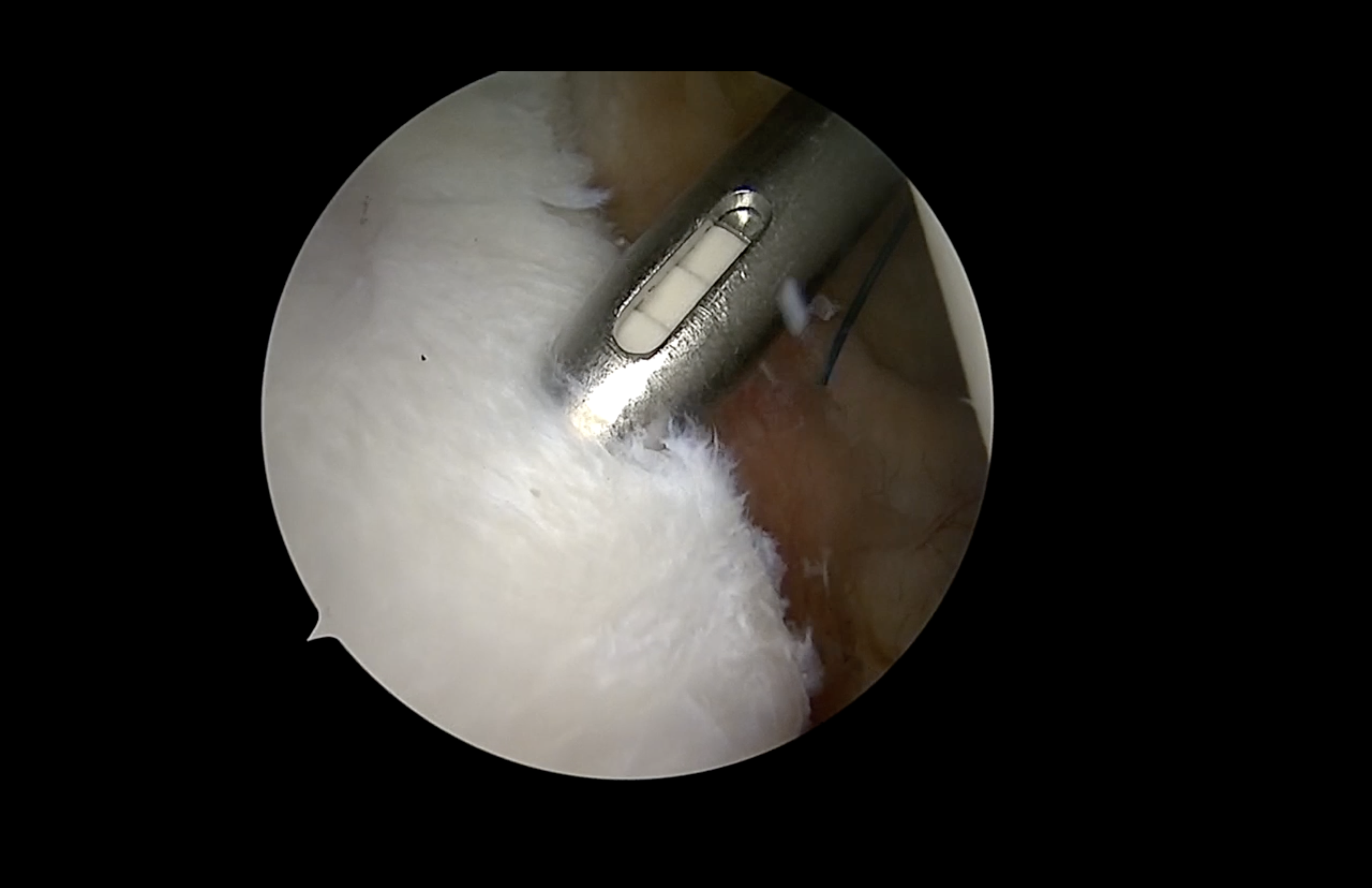
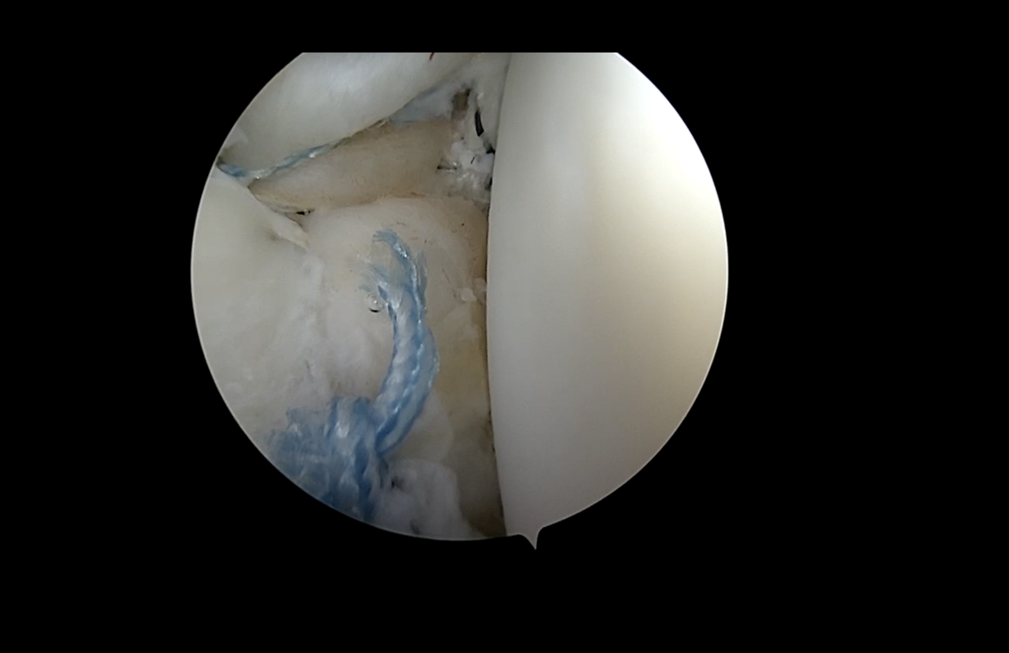
Figure 6 (A–B). (A) Anchor insertion on the anterior aspect of the labrum for Bankart lesion repair. (B) Completed standard Bankart capsulolabral reconstruction. -
Subacromial visualization.
Redirect the arthroscope to the subacromial space. Perform only a minimal bursectomy to identify the sutures. Avoid contact between the shaver and the suture limbs.
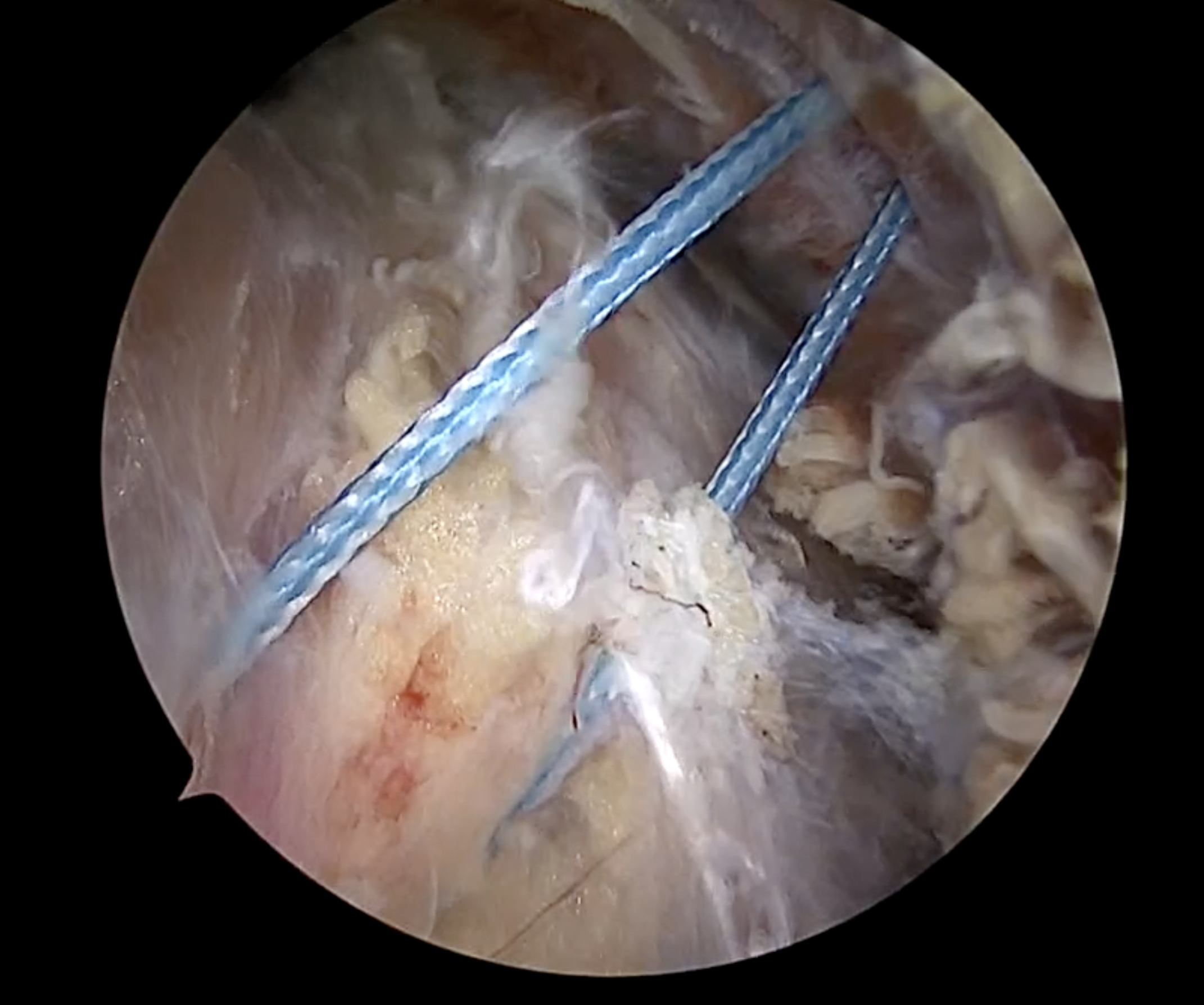
Figure 7. Subacromial visualization after minimal bursectomy to identify the passed suture limbs. -
Suture identification and tying.
Create a 2-mm punctiform portal anterior to the accessory portal. Shuttle each stitch individually and confirm that no strands are crossed or bridged. Tie the sutures through the accessory posterior portal, securing the superior oblique stitch first, followed by the posteroinferior stitch, taking care to avoid suture bridging between the two.
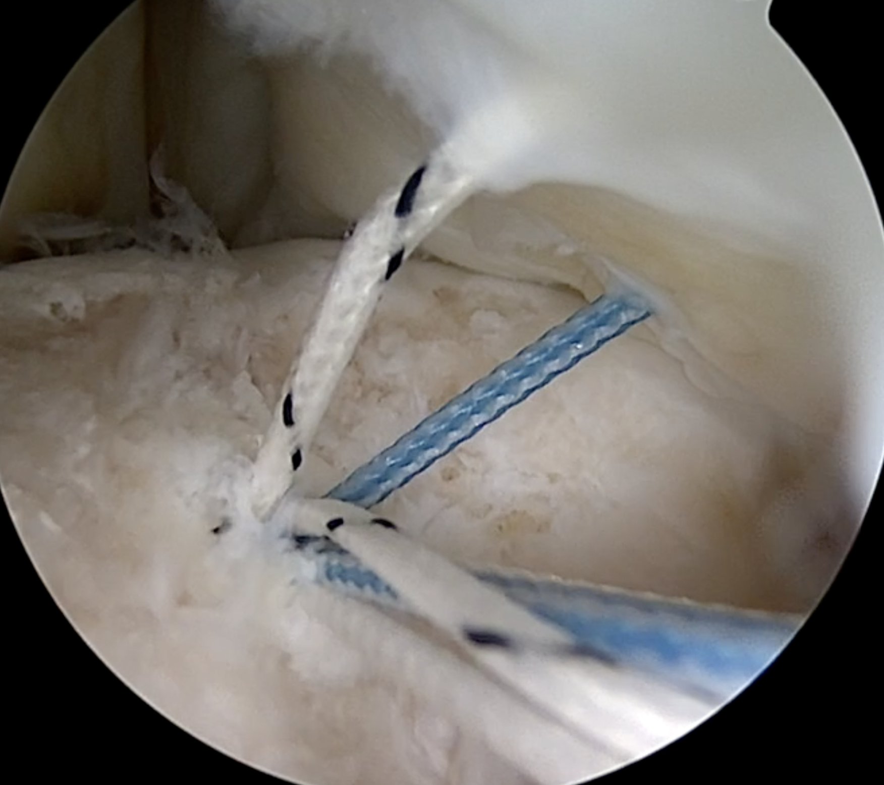
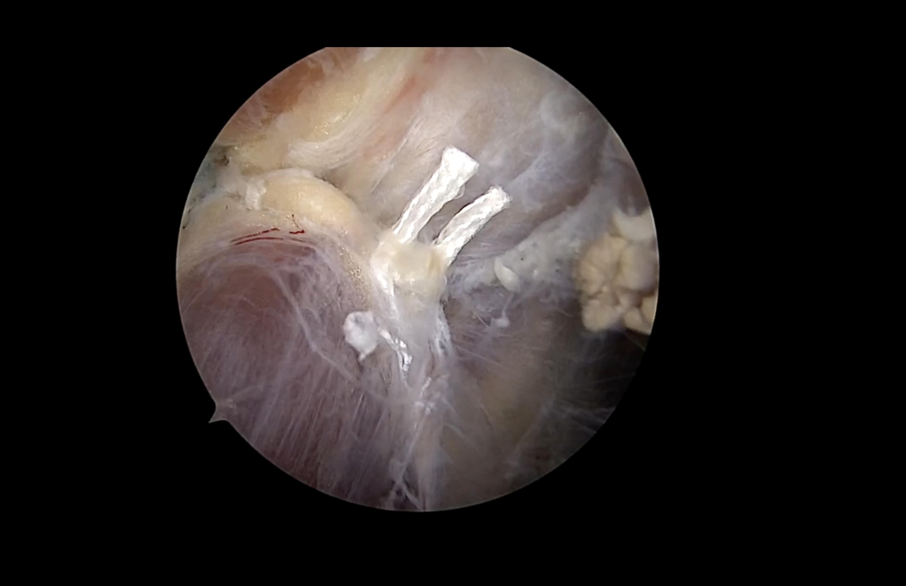
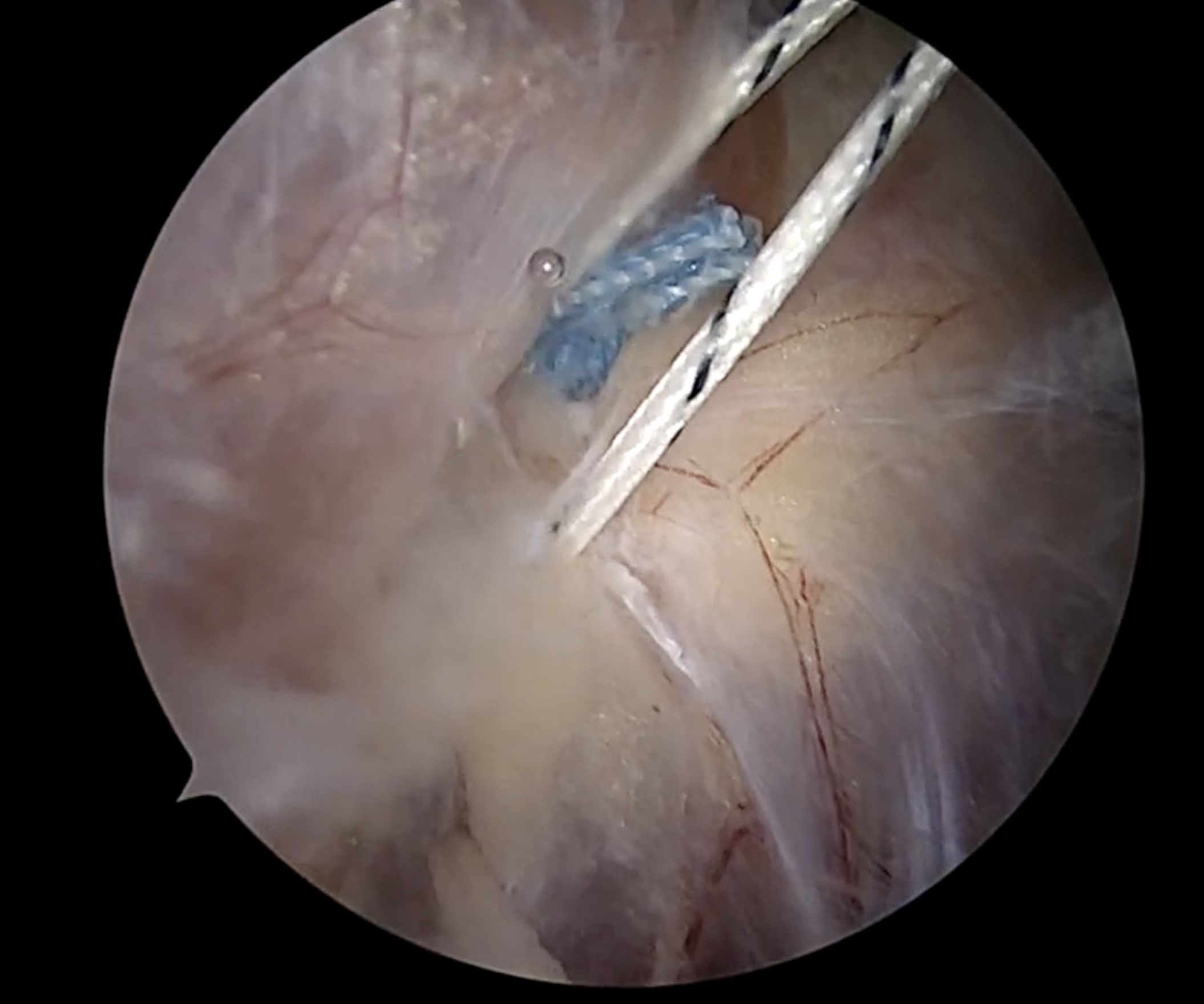
Figure 8. A: Intra-articular view of the individually shuttled stitches. B: Subacromial view of the shuttled posteroinferior stitch prior to tying. C: Subacromial view of the secured superior oblique knot. D: Subacromial view confirming the posteroinferior knot alongside the superior oblique stitch, without suture bridging.
Technical Pearls and Pitfalls
| Technical point | Purpose |
|---|---|
| Preserve a solid bony bed | Improves anchor fixation and reduces the risk of cartilage injury. |
| Identify every suture individually | Prevents bridging, crossing, knot stacking, and loss of sliding. |
| Use direct subacromial visualization | Allows confirmation of tissue capture and knot security. |
| Adjust the capsular bite to the measured laxity | Reduces the risk of residual laxity or excessive stiffness. |
Potential Advantages
The technique is intended to increase soft-tissue coverage of the Hill-Sachs defect while adding a posteroinferior capsular restraint. The use of two anchors may allow broader coverage in larger defects. The third, posteroinferior stitch is also designed to act as a mechanical backup, maintaining capsular tension and construct integrity should the deep central or superior oblique stitch fail.
These proposed advantages are based on the mechanical design of the construct and should not be interpreted as evidence of clinical superiority. Existing remplissage studies have demonstrated favorable functional and stability results, with lower recurrence than isolated Bankart repair and outcomes comparable to the Latarjet procedure at lower complication rates [9].
Limitations and Risks
- Technical difficulty and a potentially longer learning curve, as the surgeon must manage up to 12 filaments through a single portal
- Increased operative time
- Additional accessory and punctiform portals
- Risk of cartilage injury during anchor placement
- Potential suture damage during subacromial instrumentation
- Postoperative pain
- Loss of external rotation
- Postoperative stiffness caused by excessive capsular tightening
Discussion
The Scorpion/Spider technique is based on the premise that selected cases of recurrent instability contain both a structural humeral defect and a soft-tissue capsular abnormality. Treating only the anterior labrum may not fully address instability when excessive capsular volume permits translation in more than one direction.
The on-track/off-track model provides a useful framework for treatment planning because it evaluates the dynamic relationship between bipolar bone defects rather than considering the Hill-Sachs lesion in isolation [10]. This assessment remains important when deciding whether a soft-tissue procedure is appropriate or whether glenoid augmentation should be considered.
Conventional remplissage limits contact between the Hill-Sachs lesion and the anterior glenoid rim by incorporating the posterior capsule and infraspinatus tendon into the defect. The current technique preserves this principle but adds a posteroinferior capsular-shift stitch intended to reduce capsular redundancy on the humeral side, echoing the volume-reduction goal of the classic open inferior capsular shift while remaining fully arthroscopic [6].
The deep central stitch is designed to maximize compression within the defect. The superior oblique stitch extends coverage across the superior and anterior margins, while the posteroinferior stitch addresses a separate component of capsular laxity. The construct therefore provides a multi-vector repair rather than a single line of fixation, and together the three stitches are intended to create a solid, rigid remplissage that secures the joint in over 360 directions.
The clinical relevance of this design may be greatest in competitive athletes. Off-track Hill-Sachs lesions have been associated with markedly increased recurrence after arthroscopic Bankart repair [11]. However, athletic participation alone should not determine treatment. Sport type, age, instability direction, bone loss, capsular laxity, previous surgery, and the patient's expectations should all be considered.
The technique remains fundamentally different from the Latarjet procedure. Latarjet increases anterior glenoid width and provides a dynamic sling, whereas the Scorpion/Spider technique uses soft-tissue fixation without bone transfer. The construct is designed with the goal of approaching a recurrence rate comparable to Latarjet in appropriately selected patients without critical glenoid bone loss; whether it in fact achieves an equivalent recurrence rate has not yet been demonstrated and requires direct comparative data.
Postoperative motion is an important concern. Incorporation of the infraspinatus and posterior capsule may restrict external rotation, while the additional capsular shift could increase this effect if excessive tissue is captured or overtensioned. Comparative evidence has not consistently demonstrated a statistically significant difference in external-rotation loss between remplissage, Bankart repair, and Latarjet [12]. Nevertheless, motion should be measured prospectively for this specific construct.
Accurate anchor trajectory, individual suture shuttling, and cautious subacromial instrumentation are essential to avoid the technical risks summarized above. The present description is a technical proposal and does not establish clinical superiority; claims regarding lower recurrence, earlier rehabilitation, return to competitive sport, or outcomes equivalent to Latarjet require comparative clinical data.
Future studies should define patient selection using reproducible imaging and clinical criteria, including glenoid bone loss, Hill-Sachs interval, glenoid-track status, lesion orientation, generalized laxity, multidirectional translation, previous stabilization, sport type, and patient age [7]. Recommended outcomes include recurrent dislocation, subluxation, apprehension, revision surgery, validated shoulder scores, external rotation, strength, return to sport, complications, and imaging evidence of healing. A prospective comparison between standard Bankart repair with remplissage and the Scorpion/Spider technique is needed to determine whether the additional capsular shift provides a meaningful clinical benefit.
Postoperative Rehabilitation
Rehabilitation should be individualized and supervised by the operating surgeon and an experienced physical therapist, with early protection of the capsulotenodesis and capsular shift balanced against early initiation of rotational motion. The general framework used following this construct is summarized below.
| Phase | Timeline | Details |
|---|---|---|
| Immobilization | Weeks 0–4 | Sling immobilization in neutral rotation to protect the multi-stitch capsulotenodesis. |
| Passive ROM | Weeks 0–4 | Passive forward elevation to 120°; external rotation restricted to 0° (neutral). |
| ROM progression | Weeks 4–12 | External rotation advanced to 30° by week 6. Active ROM begins at week 6, progressing to full, unrestricted ROM by week 12. |
| Strengthening | Weeks 6–8 onward | Submaximal isometrics from week 6; progressive isotonic strengthening of the rotator cuff and periscapular musculature from week 8. |
| Return to sport | Months 4–6+ | Non-contact sports at 4–5 months. Contact or overhead sports at 6+ months, requiring full painless ROM, complete absence of apprehension, and >90% strength symmetry on isokinetic testing. |
Conclusion
The Scorpion/Spider technique combines a three-stitch remplissage with a posteroinferior humeral-sided capsular shift. It is designed for carefully selected patients with anterior instability, a Hill-Sachs defect, and multidirectional capsular laxity. The technique may offer a joint-preserving option in patients without critical glenoid bone loss, but its safety and effectiveness require clinical validation.
Disclosures
Informed Consent: Informed consent was obtained from the patient(s) for the use of intraoperative images and video, and for the publication of this report.
Conflict of Interest: The authors declare that they have no conflict of interest relevant to this work.
Funding: This study received no funding from any public, commercial, or not-for-profit funding agency.
References
- Provencher MT, Frank RM, Leclere LE, et al. The Hill-Sachs lesion: diagnosis, classification, and management. J Am Acad Orthop Surg. 2012;20(4):242–252.
- Boileau P, Villalba M, Héry J-Y, Balg F, Ahrens P, Neyton L. Risk factors for recurrence of shoulder instability after arthroscopic Bankart repair. J Bone Joint Surg Am. 2006;88(8):1755–1763.
- Purchase RJ, Wolf EM, Hobgood ER, Pollock ME, Smalley CC. Hill-Sachs "remplissage": an arthroscopic solution for the engaging Hill-Sachs lesion. Arthroscopy. 2008;24(6):723–726.
- Longo UG, Rizzello G, Loppini M, et al. Multidirectional instability of the shoulder: a systematic review. Arthroscopy. 2015;31(12):2431–2443.
- Zhang M, Liu J, Jia Y, Zhang G, Zhou J, Wu D, Jiang J, Yun X. Risk factors for recurrence after Bankart repair: a systematic review and meta-analysis. J Orthop Surg Res. 2022;17(1):113.
- Neer CS 2nd, Foster CR. Inferior capsular shift for involuntary inferior and multidirectional instability of the shoulder: a preliminary report. J Bone Joint Surg Am. 1980;62(6):897–908.
- Gaskill TR, Taylor DC, Millett PJ. Management of multidirectional instability of the shoulder. J Am Acad Orthop Surg. 2011;19(12):758–767.
- Itoi E. "On-track" and "off-track" shoulder lesions. EFORT Open Rev. 2017;2(8):343–351.
- Hurley ET, Toale JP, Davey MS, Colasanti CA, Pauzenberger L, Strauss EJ, Mullett H. Remplissage for anterior shoulder instability with Hill-Sachs lesions: a systematic review and meta-analysis. J Shoulder Elbow Surg. 2020;29(12):2487–2494.
- Di Giacomo G, Itoi E, Burkhart SS. Evolving concept of bipolar bone loss and the Hill-Sachs lesion: from engaging/non-engaging lesion to on-track/off-track lesion. Arthroscopy. 2014;30(1):90–98.
- Shaha JS, Cook JB, Rowles DJ, Bottoni CR, Shaha SH, Tokish JM. Clinical validation of the glenoid track concept in anterior glenohumeral instability. J Bone Joint Surg Am. 2016;98(22):1918–1923.
- Gonzalez-Morgado D, Ardebol J, Noble MB, Galasso LA, Menendez ME, Denard PJ. No difference in external rotation loss after isolated Bankart repair, remplissage, or Latarjet: a systematic review and meta-analysis. Am J Sports Med. 2025;53(2):493–500.
Before submission, verify the complete bibliographic details against the target journal's reference style, and confirm figure/video legend formatting requirements.